INTRODUCTION
In the craniofacial area, osteosarcoma frequently affects jaw bones with approximately 6% of osteosarcomas of the mandible or maxilla. It tends to occur approximately in people older than 40 years that is a decade later than osteosarcomas of the long bones [1]. Males are affected more frequently than females.
Osteosarcomas usually occur as secondary tumors after radiation therapy or chemotherapy administered for a preexisting tumor [2]. In a pediatric patient without a previous history of irradiation to the head and neck area, a primary osteosarcoma of the turbinate is extremely rare. To the best of our knowledge, a primary osteosarcoma arising from the middle turbinate has not been reported in the English literature. We also reviewed the previously reported cases of tumor arising from turbinate.
CASE REPORT
A 9-year-old girl was admitted with flu like symptoms and progressive nasal obstruction. She didn't complain of epistaxis and had no history of surgery or facial trauma. The results of other routine laboratory examinations were normal. Physical examination showed fullness, convexity and lateral displacement of left nasal bony area.
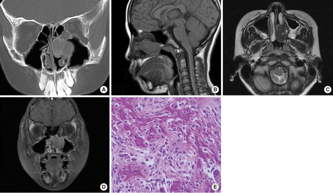
Non-contrast computed tomography (CT) examination revealed a 1.5×3.0×2.0-cm well-defined hyperattenuating mass, which originated from the middle turbinate (Fig. 1A). Within the mass, there was loss of turbinate detail and no evidence of calcification. On magnetic resonance imaging (MRI) examination, the mass revealed intermediate signal intensities on T1-weighted images (WI) (Fig. 1B) and low signal intensities with focally high signal intensities on T2-WI (Fig. 1C). The mass was enhanced by injection of gadopentetate dimeglumine (Fig. 1D).
The mass was removed by endoscopic sinus surgery under general anesthesia. The mass was a mucosa-covered fixed, and hypervascular solid mass. During the intraoperative or postoperative period, excessive bleeding did not detected. Both intraoperative frozen section margins and final pathologic margins (anterior wall and lateral wall of sphenoid, adenoid, anterior and posterior ethmoid, fontanelle, superior wall of middle turbinate, agger nasi cell, and sphenopalatine foramen) were negative. There was no histologic evidence of tumor invasion to surrounding sinus walls or basal lamella of middle turbinate. A pathological examination of the mass identified the presence of an osteoblastic osteosarcoma, grade II/III (Fig. 1E). On the immunohistochemical staining, the mass is negative for c erb B2 and positive for RB gene protein and amount of Ki-67-positive cells is approximately 15% in cross section.
After surgery, she was relieved of any symptoms and nasal stuffiness. She has been followed up for 1 year without any evidence of disease recurrence.
DISCUSSION
Only nine cases of turbinate tumors have been previously described in the literature if osteomas arising from turbinate are ruled out, to our knowledge (3-12). Most of them were benign tumor and only three cases of them were malignant tumor. Radiologically, bone destruction which is a reliable sign of malignancy was observed in one case of malignant tumors. These findings can't be useful in determination of whether the mass is benign or malignant. Clinical symptoms were variable but nasal obstruction was most common presenting symptom (Table 1).
A few cases of turbinate osteomas have been reported. And the main symptoms were headache or facial pain. These tumors showed calcifications or calculus within the mass on CT scans which is useful clue to the diagnosis.
Enlargement of the tumor can obstruct sinus ostium and lead to mucocele formation or infection. Unilateral obstruction is the most common presenting symptom of a nasal tumor but epistaxis, rhinorrhea, and sinusitis may also be present. Diagnosis can be delayed, because the signs and symptoms of that are similar to benign sinonasal disease. Moreover, because of the low incidence of turbinate tumor, we tend to ignore the possibility of neoplasm. If a physician has a high index of suspicion, CT scanning can be the study of choice and endoscopic biopsy should be done to rule out the possibility of neoplasm.
In general, a benign turbinate tumor is a well-defined, localized mass without remodeling or thickening of adjacent bone whereas a malignant tumor is an ill defined, bony destructive mass. But a bony destructive mass is not pathognomic sign of malignancy because benign tumors may also appear quite aggressive.
CT and MRI scans are effective methods for specification of the anatomic site of the nasal airway obstruction. Imaging findings of malignant turbinate tumors could reveal loss of turbinate detail, mineralization, invasion of surrounding bone, mass-like soft tissue mass, contrast uptake, and area of high attenuation on CT. A hyperattenuating mass might be seen due to the presence of calcium and/or traces of metallic element (13).
It is conceivable that inverted papilloma or fibro-osseous lesion such as fibrous dysplasia can occur in the turbinate. Inverted papilloma often is coincident with calcification and adjacent inflammatory mucosa. Fibrous dysplasia appears ground glass like lesion. Fungal rhinitis may appear as mass-like lesion and may mimic a malignancy with bony destruction. The presence of ferromagnetic element or tight hydrogen binding will helpful to the diagnosis of fungal sinusitis. It causes the finding of hypointensity on T1-WI and markedly hypointensity on T2-WI (14).
In conclusion, we report here on rare case of primary turbinate osteosarcoma in a 9-year-old girl without radiation therapy or chemotherapy, presenting as a unilateral hyperattenuating soft tissue mass with loss of turbinate detail on non-contrast CT and enhancing mass on MRI with intermediate signal intensity on T1-WI, low signal intensity on T2-WI.