INTRODUCTION
Myringosclerosis is an irreversible pathologic healing mechanism of the tympanic membrane which can result in sclerotic plaques of the membrane. Myringotomy and tympanic membrane perforation due to otitis media can result in myringosclerosis. The pathologic process of the myringosclerosis is thought to be a part of hyperoxia as the trigger of producing free radicals from the inflamed tissue and observed as deposition of calcareous plaques in the fibrous layer of the tympanic membrane [1,2]. Antioxidant treatment is a well-known prevention therapy and, during the last decade, different groups of antioxidants were used in this manner [3,4,5,6,7]. These treatments are mostly experimental but antioxidant agents have been used for human trials as well [8]. The primary means of determining the benefit of antioxidant agents during the sclerotic stage is by otomycroscopy, biochemistry and histopathologic examination.
Coenzyme Q10 (CoQ10) is a lipid soluble antioxidant which is synthesized endogenously in our bodies. It has an important role in mitochondrial electron transfer and energy conservation [9]. However, it is a very effective antioxidant that protects the cellular membrane from free oxyradical formation [10].
Lycopene, which is responsible for yellow, orange or red pigmentation, is present in tomatoes and other red fruits and vegetables and is the most prevalent carotenoid in the Mediterranean diet. Several experimental rodent studies suggest that lycopene possesses many biochemical functions, including inhibition of proinflammatory activities, antioxidant scavenger and protrombotic factors [11]. Additionally, it exerts a critical role in the inflammatory process and can decrease lipid peroxidation, increase neutrophil infiltration and apoptosis [12,13].
Grape seed extract (GSE) contains proteins, lipids, carbohydrates, and natural antioxidant proanthocyanidins, which have been demonstrated to have an effect against oxidative stress and free radicals [14,15].
This study was designed to determine the effect of CoQ10, lycopene, and GSE. The aim of this study is to compare the effectiveness of these antioxidants by examining the tympanic membranes, both otomicroscopically and histopathologically.
MATERIALS AND METHODS
The study complied with experimental ethical principles and animal protection laws according to the rules and regulations in Turkey, approved by the Local Ethic Committee in Ankara, Turkey (Prot. No.: 20;15/07/2011). All animal care and procedures were performed humanely. The animals were kept in ordinary cages (15 cm├Ś25 cm├Ś40 cm) and were fed with standard rat food and tap water. Temperature was 23.3Ōäā with artificial lighting in a period of 12 hours. The rats were used after 10 days of quarantine.
Animals and procedures
Fifty Wistar-Albino male rats weighing 340-450 g were used for the procedure and each was examined to confirm an intact tympanic membrane otomicroscopically (Opmi 1, Carl Zeiss, Munich, Germany). Two animals with impacted cerumen, which could not be removed, were excluded from the study. Forty-eight animals were anesthetized with 80-mg/kg ketamine hydrochloride. Under otomicroscopic examination with a sterile pick, bilateral myringotomies were performed on the posterosuperior eardrum by the two senior authors (SA and AT). After the procedure, four animals were excluded from the study due to middle ear infections and intractable vertigo.
Following myringotomy, the animals were randomly divided into four groups. Group 1 (saline) comprised of 11 rats to which 2 mL/kg daily saline was administered orally as the control group. Group 2 comprised 11 of rats to which a daily dose of 100-mg/kg grape seed extract (GSE) (GNC standardized grape seed extract 300 mg/100 capsules) in a 2-mL solution was administered orally. Group 3 comprised of 11 rats to which a daily dose of 100-mg/kg CoQ10 (GNC CoQ10 100 mg/30 capsules) in a 2-mL solution was administered. Lastly, group 4 comprised of 11 rats to which a daily dose of 200-mg/kg lycopene (GNC lycopene from tomatoes 30 mg/60 capsules) in a 2-mL solution was administered orally, as well. All drugs, including saline, were administered orally for 14 days starting on the same day of surgery.
Otoscopic examination
All rats were anesthetized with 80-mg/kg ketamine hydrochloride at the end of the 14th day. Firstly, otomicroscopic examinations were performed by the two senior authors (SA and AT) randomly, without knowing each other's findings or the groups to which the rats belonged. Tympanic membrane lesions were evaluated by a four point scale, described by Mattsson et al. [1]; 0, no visible myringosclerosis (MS); +, occasional MS (sclerotic lesion was present around umbo); ++, moderate MS (sclerotic lesion was present around umbo) and beside the handle of the malleus and along the anterosuperior portion of the pars tensa; +++, MS sclerotic lesion was present on the malleus and along annulus. The rats were euthanized by thiopental sodium injection and then their tympanic bullae were set aside for histopathological examination.
Histopathological examination
Specimens were evaluated by one pathologist who was blinded to the groups and all specimens were prepared as follows.
The tympanic membrane (TM) and surrounding bony annuli were removed together by microdissection under otomicroscopy. Before fixation of specimen myringotomy regions of the specimens are visualized with small amount of tissue marker. All specimens were routinely fixed with 10% buffered formalin overnight and decalcified with 10% formic acid. After decalcification specimens were dehydrated with graded alcohol baths and sampled including the myringotomy region. Paraffin-embedded blocks were serially sectioned at 4-┬Ąm thickness, and stained with hematoxylin-eosin and Masson's trichrome stain. Masson's trichrome stain was used to visualize the fibrotic change of lamina propria.
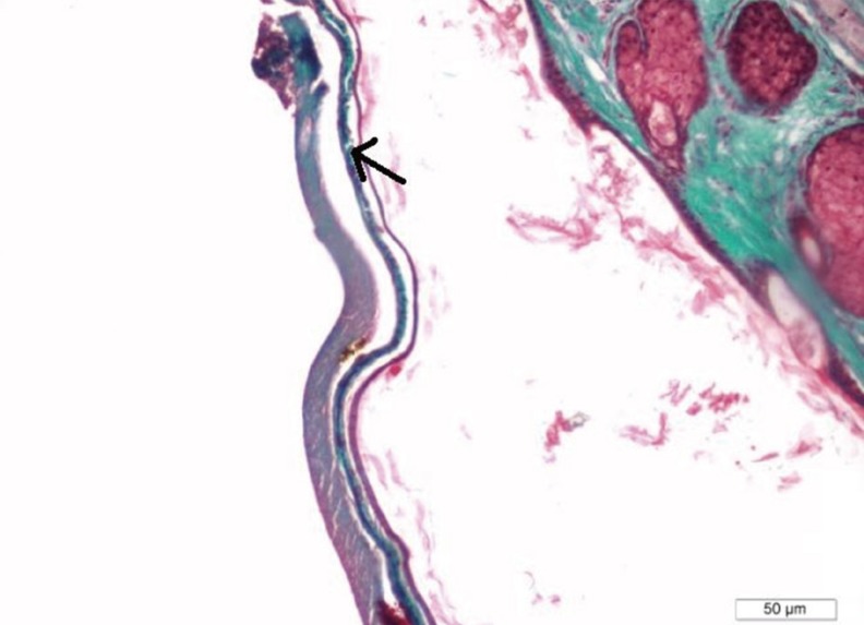
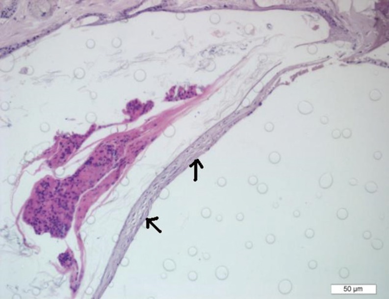
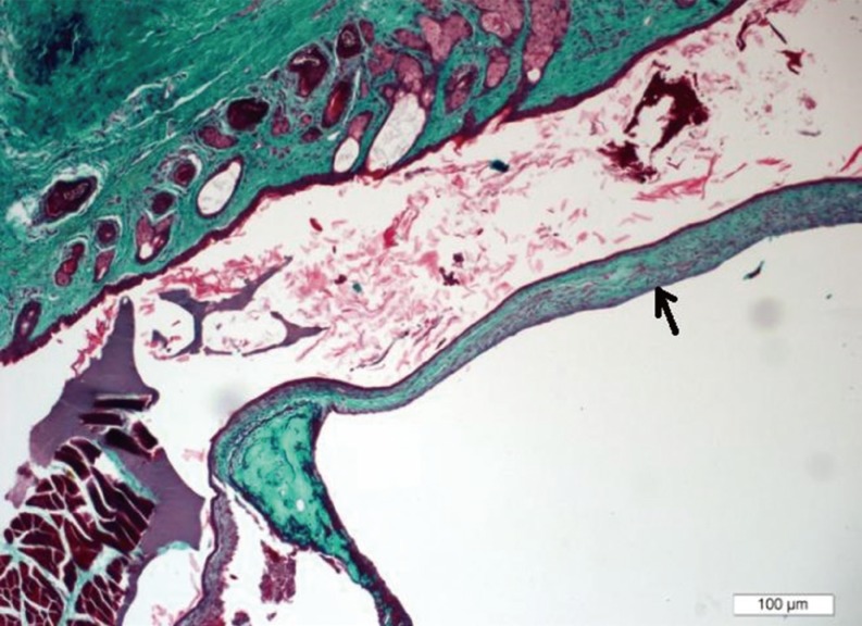
All serial sections were examined with a light microscope (DP2-BSW, Olympus, Tokyo, Japan). The scoring of the lesions was done on the section that demonstrates the maximum fibrotic change for each rat. The degree of sclerotic lesions and intensity of fibroblastic proliferation in the lamina propria of timpanic membranes were graded semiquantitatively as: 0, no visible myringosclerosis, lamina propria was free of sclerotic lesion (Fig. 1); 1, occasional miringosclerosis, few single lesions in the lamina propria (Fig. 2); 2, moderate myringosclerosis, single and confluent lesions observed in the lamina propria (Fig. 3); 3, serious myringosclerosis, extensive confluent lesions observed in the lamina propria (Figs. 4, 5) [1].
Analyses
In order to identify any statistical significance in the results, data analysis was performed using SPSS ver. 11.5 (SPSS Inc., Chicago, IL, USA). Ordinal data were shown as median (min-max). Whether or not the differences in degree of otoscopic and histopathologic examinations among groups were statistically significant was evaluated with the Kruskal-Wallis test. When the P-value from the Kruskal-Wallis test was statistically significant, the Conover multiple comparison test was applied. The Conover multiple comparison test was used to distinguish which groups differed from the other groups. A P-value less than 0.05 was considered statistically significant.
RESULTS
Based on otoscopic findings on the 14th day, all the tympanic membrane perforations were closed on all 44 rats. Comparison of the saline group against the GSE, lycopene, and CoQ10 groups revealed statistically significant differences with P<0.001. There were no statistically significant differences between GSE and lycopene, GSE and CoQ10, and lycopene and CoQ10 groups comparisons showing P-values of 0.355, 1.000, and 0.355, respectively (Fig. 6).
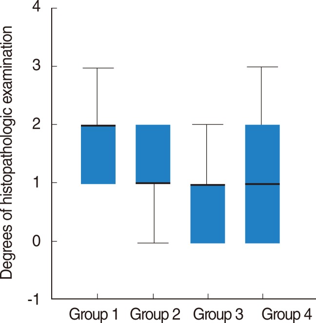
Sclerosis of tympanic membrane evaluation in histopathologic examination revealed a remarkably higher difference in the saline group compared with GSE, lycopene, and CoQ10 groups, with P-values 0.018, 0.003, and <0.001, respectively. There were no statistically significant differences between GSE and lycopene, GSE and CoQ10, and lycopene and CoQ10 groups comparisons with P-values 0.513, 0.205, and 0.536, respectively (Table 1). They were each compared against saline group and demonstrated similar antioxidative and anti-inflammatory effects.
DISCUSSION
In this study, three different kinds of antioxidants with placebo (saline) on myringotomised rat ears were compared via otoscopic examination and histopathologic findings. The objective was to evaluate the effectiveness of certain antioxidants by examining the tympanic membranes, both otomicroscopically and histopathologically. Our results revealed a successful outcome with all three antioxidants. These results are similar to other antioxidants which have been studied in the literature. However, three different antioxidants were not compared in this way before.
There are the increase of hyaline degeneration, collagen tissues and calcification in TM when MS occurred, and the real cause and pathogenesis of it are not well-known [1,16].
It can be seen that MS is an irreversible and nonspecific result of the production of free radicals which occurred after ventilation tube (insertion) or trauma to the TM [3,16,17].
Tympanic membrane lesions were evaluated by a 4-point scale previously described by Mattsson et al. [1], which was used as the primary way to demonstrate the effects of three different antioxidant antioxidant agents in this study.
Reviewing the literature, experimental studies including different kind of antioxidant agents for preventing myringosclerosis shows success promising results. Ozcan et al. [4] topically applied two different dosages of N-acetylcysteine (NAC) in myringotomised rat ears and found this had a significant effect on preventing sclerotic lesions using otomyroscopy and histopathology on both groups. However, there were no significant differences between the two NAC groups. Song et al. [18] studied the effects of caffeic acid phenethyl ester (CAPE) on rats and their results indicated that, based on histopathologic exam, CAPE is a preventive antioxidant. Furthermore, Gorur et al. [19] administered selenium systematically to the myringotomised rats and found statistically significant lower tympanosclerosis than in the control group. The results of this study showed that when compared against the saline group, there were statistically significant differences with sclerosis degrees in GSE, lycopene, and CoQ10 groups <0.001.
In similar experiments, the topical application of ascorbic acid on perforated rat ears revealed significant difference between saline treated (untreated) and ascorbic acid treated groups, with the ascorbic acid demonstrating positive results [6].
Uneri et al. [8] used topical vitamin E on seventy eight childrens' right ears just after bilateral ventilation tube insertion and observed the subjects for 9 months. Their results indicated that vitamin E treated ears had a significant improvement compared to the untreated ears.
Mattsson et al. [1] hypothesized that a hyperoxic condition (existed) in the middle ear cavity and the inflammation induced by tympanic membrane trauma and inflammatory reaction consisting of inflammatory cells around sclerotic aggregates, resulting in an increased production of O2-derived free radicals, may be the cause of MS pathogenesis. It has been proven through experimental models that a relationship exists between O2-derived radicals and the occurrence of MS. It was also shown that the application of various free radical scavengers, antioxidants and anti-inflammatory agents could reduce the formation of MS after experimental myringotomy [1,6,7,18,19].
CoQ10 is a molecule that protects the cellular membrane from free oxyradical formation [10]. Additionally, this molecule has an important role in mitocondrial electron transfer and energy conservation [9]. The CoQ10 molecule inhibits lipid peroxidation by preventing the production of lipid peroxyl radicals (LOO) and reduces the initial perferryl radical and singlet oxygen, with concomitant formation of ubisemiquinone and H2O2. This is achieved because the molecule is continually going through an oxidations-reduction cycle. CoQ10 protects lipids and proteins from oxidation by quenching of the initiation perferryl radicals.
Lycopene is an inhibitor of proinflammatory, antioxidant scavenger and protrombotic factors [11]. Lycopene also exerts a critical role in the inflammatory process and can decrease lipid peroxidation, increase neutrofil infiltration and apoptosis [12,13]. Lycopene, however, does not have a beta-ioning ring structure, which subsequently increases the molecules antioxidant action. The presence of lycopene in the Ames test can trap singlet oxygen and reduce mutagenesis. Inhibition of the formation of thiobarbituric acid-reactive substances is directly related to the antioxidant activity of carotenoids in multilamellar liposomes.
An effective treatment for oxidative stress and free radicals is GSE [14,15]. The reason for this is because GSE contains large amounts of phenolic compounds which are responsible for antioxidant activity. GSE can reliably indicate antioxidant activity by determining whether or not antioxidant compounds are electron donors. In turn, this can reduce the oxidized intermediates of the lipid peroxidation process [20].
In conclusion, the formation of myringosclerotic plagues after experimental myringotomy in rats significantly decreased and diminished after systemic administration of CoQ10, lycopene, and GSE, which have been demonstrated to have an effect against oxidative stress and free radicals. They were compared against a saline group and their antioxidative and anti-inflammatory effects were similar.
Prevention and treatment effects of CoQ10, lycopene, and GSE may be useful for humans who are at risk of developing myringosclerosis and tympanosclerosis. Therefore, more elaborate clinical research should be designed and undertaken to confirm this expectation.