INTRODUCTION
The lingual artery is distributed in base of tongue and mobile tongue and susceptible damage during operation. Such injury can lead to immediate or delayed hemorrhage, which can be life threatening. If both lingual arteries are damaged, necrosis of mobile tongue below damaged point is inevitable. Knowledge of anatomical distribution of the lingual artery is crucial during surgery such as partial glossectomy or tongue base surgery to reduce complications. It is also helpful to decide resection margin and reconstruction.
The lingual artery originates from the external carotid artery just above the superior thyroid artery and runs into the intrinsic muscle of tongue through the posterior border of the hyoglossus muscle. The lingual artery gives first branch, dorsal lingual artery, while passing the hyoglossus muscle. It proceeds anterosuperiorly and gives sublingual artery at the anterior border of hyoglossus muscle. After giving sublingual artery, the lingual artery shifts to deep lingual artery. Deep lingual artery runs through inferior portion of mobile tongue giving multiple perforating branches toward dorsum of the tongue. The lingual artery can be subdivided into 4 segments. The first segment is original segment which starts from hyoid bone to the point where dorsal lingual artery is given. The 2nd part, hyoglossus segment and the 3rd part, ascending segment is divided where the sublingual artery originates. Deep lingual artery is designated as horizontal segment [1].
There were several previous studies to analyze the course of the lingual artery and the anatomical markers. Hou et al. [2,3] used computed tomographic angiography to analyze anatomic relationship between the lingual artery and lingual markers and to set safety space for functional surgery on the tongue. Kimura et al. [4] used Doppler sonography to evaluate the lingual artery at the midpoint of tongue. Lopez et al. [5] studied about vascular territory of the lingual artery using selective ink injections in 15 fresh cadaver heads. But none of these studies researched histologic features of the lingual artery on mobile tongue. In this study, we made 18 healthy cadaveric tongues into 8 coronal sections each to clarify the anatomical distribution of the lingual artery in normal Korean subjects by light microscopic observation.
MATERIALS AND METHODS
Subjects
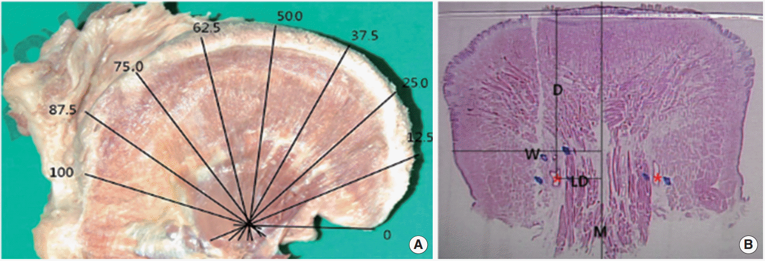
For this study, eighteen healthy cadaveric tongues were used to get 8 pieces of paraffin-embeded tissue section each. Fifteen cases were male, 3 cases were female. Median age was 62 years old ranging from 31 to 88 years. Before producing histologic section, we chose one specimen and performed intramuscular dissection of the tongue to identify the course of the lingual artery. The neck has been partially dissected by medical students and further dissections were performed by the authors. After identifying external carotid artery, we could find the lingual artery just above the superior thyroid artery at the level of hyoid bone. We removed hyoglossus muscle to expose the lingual artery and careful intramuscular dissection was done along the artery. The lingual artery enters base of tongue at greater cornu of hyoid bone and runs along the medial surface of hyoglossus muscle. It proceeds antero-superiorly and shifts as a deep lingual artery at mobile portion of the tongue which runs along the inferior surface of the mobile tongue (Fig. 1). This specimen was not included in histologic slides. Eighteen cadaveric tongues were measured the lengths from the tip of the tongue to foramen cecum. Each tongue was divided into 8 coronal sections according to the dorsal lengths of each tongue. We designated tongue tip as a ‘0%’ spot and foramen cecum as a ‘100%’ spot and acquired tissue sections at 12.5%, 25.0%, 37.5%, 50.0%, 62.5%, 75.0%, 87.5%, and 100% spots. Tissue cutting was performed along the vertical plane of dorsal surface of the tongue (Fig. 2A).
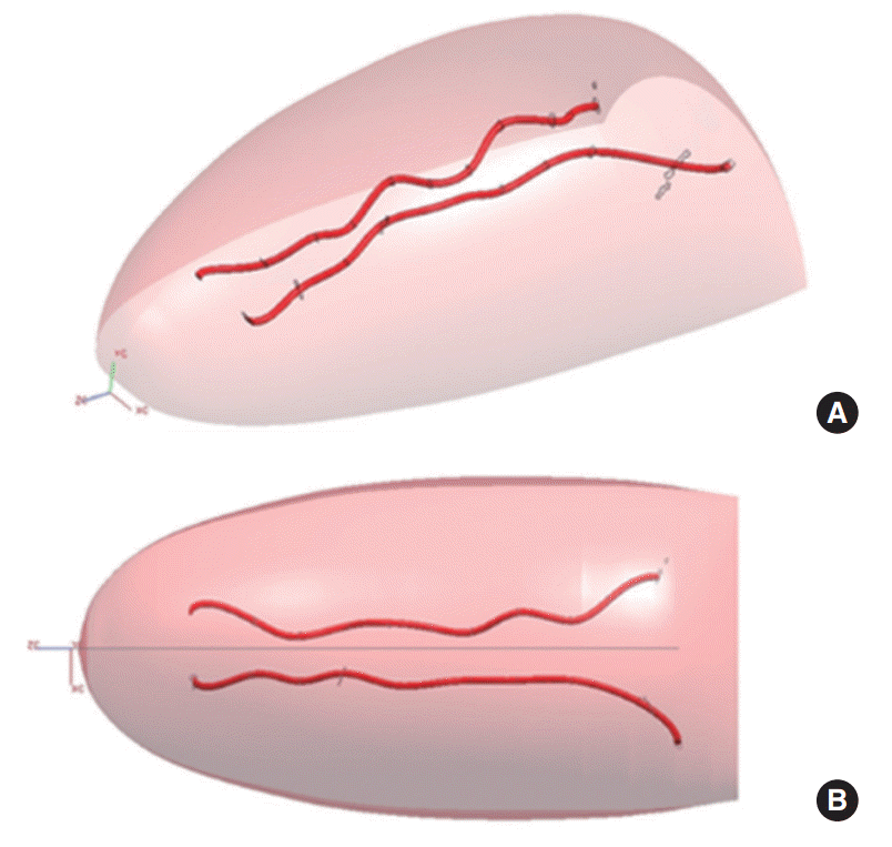
The Formalin fixed, paraffin-embedded giant tissue slides were produced and hematoxylin-eosin stains were added. The slides were examined under light microscope. The length from midline raphe, depth (D) from dorsal surface of tongue and the whole transverse length of tongue were measured using Storz E-2410 caliper (Storz, St. Louis, MO, USA) but perforating branches of the lingual artery were excluded. The lateral distance (LD), D, and proportion of LD (P) of deep lingual artery were determined from tip to base of tongue gradually. LD is length from median raphe to the center of deep lingual artery lumen. D is vertical distance from dorsal surface of tongue to the center of deep lingual artery. P is obtained by dividing LD with transverse length from median raphe to lateral border of tongue (Fig. 2B). Thereafter, the degree of symmetry between right and left side and the difference between selected zones (25%, 50%, 75%, and 100% spots from tip to foramen cecum) were evaluated. We reconstructed 3-dimensional images of lingual artery using Unigraphics ver. 2.0 (Unigraphics Solutions Inc., Maryland Heights, MO, USA) based on data from the representative case (Fig. 3).
Statistical analysis
The differences between the right and left side locations of the lingual artery were investigated by Wilcoxon sign ranks test. The differences in 25%, 50%, 75%, and 100% spots mean values of LD, D, and P were analyzed using Kruskal-Wallis test. One-to-one comparison of mean values of selected spots (which were 25%, 50%, 75%, and 100%) were performed using Wilcoxon sign ranks test. All differences were defined as significant for a P-value less than 0.05. These analyses were performed using the statistical software PASW SPSS ver. 18.0 (SPSS Inc., Chicago, IL, USA).
RESULTS
Lateral distance of the lingual artery
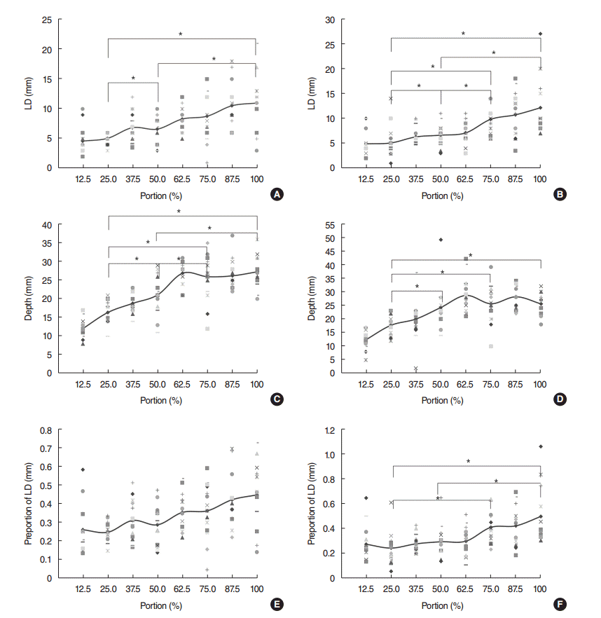
The average LD from midline raphe of the tongue were listed on Table 1. LD of 12.5% spot was the shorter than any other values (right side, 4.6±2.5 mm; left side, 4.9±2.7 mm) while LD of foramen cecum was the largest one (right side, 11.0±4.8 mm; left side, 12.2±5.9 mm). There was no significant difference between right and left side (P>0.05). LD was gradually increased as lingual artery runs toward foramen cecum (Fig. 4A, B). Statistical analysis of LD at 25%, 50%, 75%, and 100% spots using Kruskal-Wallis test showed significant difference in right and left side (P<0.05). On one-to-one comparison using Wilcoxon sign rank test, 25% spot was significantly different from 50% and 100% spots and so was 50% spot from 100% in right side (P<0.05). There was no significant difference between 25%–75%, 50%–75%, and 75%–100% spots (P>0.05). In the left side however, there were significant differences in 25%–50%, 25%–75%, 25%–100%, 50%–75%, and 50%–100% comparison (P<0.05). Only the 75%–100% spot showed no difference in the left side (P>0.05).
Depth of the lingual artery
Mean values of D were shown in Table 2. There was no difference between the right and left sides (P>0.05). Mean values of D at 12.5% spot were 12.1±2.5 mm (right) and 12.5±3.8 mm (left) and mean values of D at foramen cecum were 27.3±4.5 mm (right) and 25.5±4.2 mm (left). The lingual artery placed deeper as it run from tongue tip to the foramen cecum (Fig. 4C, D). There was significant difference in comparison between 25%, 50%, 75%, and 100% spots using Kruskal wallis test (P<0.05). On Wilcoxon sign rank test, the mean values of D were significantly different except 75%–100% comparison in the right side (P<0.05). No significant differences were observed between 50%–75%, 50%–100%, and 75%–100%, but D of 25.0% was significantly different with 50%, 75%, and 100%.
Proportion of lateral distance of the lingual artery
The mean values of P were shown in Table 3. P of 12.5% was closer to midline than other spot (right, 26%±13%; left, 27% ±15%), P of foramen cecum were 45%±18% in the right side and 50%±24% in the left side which located farthest from the midline (Fig. 4E, F). No significant differences were observed between sides (P>0.05). The average P of 25%, 50%, 75%, and 100% spots showed significant difference in Kruskal-wallis test (P<0.05). On Wilcoxon sign rank test, there were no significant difference in the right side, but in the left side, 25%–75%, 25%–100%, and 50%–100% comparisons showed significant difference (P<0.05).
We reconstructed 3-dimensional images of lingual artery using Unigraphics ver. 2.0 based on data from the representative case (Fig. 3). Both sides of the lingual artery were lateralized from the median raphe and placed deeper from the surface until they reach foramen cecum. Right and left sides of the lingual artery were symmetric.
DISCUSSION
Oral cavity is limited space so only a small amount of bleeding can disturb surgical fields. Tongue has excellent mobility and abundant vasculatures. Massive bleeding can occur when main trunk of the lingual artery is damaged and ligation is difficult due to limited surgical field [6,7]. Thus, it is important to prevent excessive bleeding by preserving the lingual artery. In this study, we used eighteen cadaveric tongues to clarify anatomical distribution of the lingual artery by light microscopic observation.
There were a few studies about anatomical distributions of the lingual artery using computed tomographic angiography, Doppler sonography and cadaveric tongue [2-5,8]. Hou et al. [2] analyzed the lingual artery to define safety space for functional surgery of the tongue using computed tomographic angiography. He proposed V-zone of the tongue which is similar with our findings [2], but most of the reports focused on base of tongue and lack an explicit description of the lingual artery morphology and topography at the level of the tongue.
We tried to define useful surgical markers to facilitating a surgeon’s ability to avoid intraoperative lingual artery injury. We used median raphe, foramen cecum, tongue tip, and dorsal surface of the tongue as anatomical markers because they were relatively constant at any position. We measured the length of the dorsal surface of the tongue from tongue tip to foramen cecum then divided it into 8 coronal sections and produced histopathologic slides. D of the lingual artery from the dorsal surface of the tongue is gradually increased until the lingual artery reaches 75% of length of tongue from tongue tip. From there, D of the lingual artery remains stable to foramen cecum. D of the lingual artery varies with the position of the tongue, so measured value itself cannot be used in surgical fields which require traction into multidirections. Operator should keep in mind that the lingual artery located deeper pathway vertically from the surface as it reaches tongue base. The lingual artery runs lateral direction until three-fourths of tongue length. LD of the lingual artery from median raphe varies with the position of the tongue, too. P remains constant during forward traction of the tongue so it is helpful in surgical fields. The lingual artery is located within 50% of transverse length of tongue from median raphe. Surgical resection that does not exceed the ranges mentioned above is relatively safe. If the tumor extends more than a half of transverse length from lateral border in one side, the risk of the lingual artery damage increases. The area between lingual arteries is relatively safe. The mean LD at foramen cecum are 11.0±4.8 mm in the right side and 12.2±5.9 mm in the left side. This is consistent with Hou et al. [2]’s findings that surgery performed within approximately 15 mm of the tongue midline is relatively safe. Both sides of the lingual artery run symmetric and there is no transseptal anastomosis between sides like Houseman et al. [9]’s findings. However, the limitation of this study is that tissue shrinkage can occur during formalin fixation and microscopic slide production. The degree of shrinkage can differ according to contractility of the tissue. Tongue tissue can shrink due to intrinsic muscle’s contraction [10].
In conclusion, the lingual arteries of both sides are symmetric and run toward inferior and lateral directions from tongue tip to 75% of total length, but there is no significant difference around base of tongue. Finally the lingual artery located within 50% of transverse length of tongue from midline raphe. Lingual artery is usually sacrificed during resection of lesion involving tongue base. Distal injury of the lingual artery can be salvaged but proximal injury cause disaster in half portion of the mobile tongue. These findings provide surgeons useful information to set safe surgical margin that does not compromise function of the tongue during oral and base of tongue related surgery. The results of present study are notable because this is the first research to evaluate the histologic features of lingual artery on mobile tongue.
HIGHLIGHTS
▪ Anatomy of the lingual artery was examined in 18 healthy human cadaveric tongues.
▪ The artery in right and left side was symmetric.
▪ The artery was positioned laterally and then go deeply posterior to the base of tongue.
▪ The artery had no communication between right and left sides and was located within 50% from median raphe.