| Home | E-Submission | Sitemap | Contact Us |  
Clinical and Experimental Otorhinolaryngology > Volume 2(1); 2009 > Article
Park, Han, Lee, Han, Choung, and Park: Congenital Stapes Anomalies with Normal Eardrum

Abstract

Objectives

A non-progressive and conductive hearing loss with normal eardrum, but no history of trauma and infection, is highly suggestive of a congenital ossicular malformation. Among ossicular anomalies, stapes anomaly is the most common. The purpose of this study is to describe patterns of stapes anomaly and to analyze its surgical outcome with special reference to its patterns.

Methods

We conducted a retrospective case review. The subjects comprised 66 patients (76 ears) who were decisively confirmed by the exploratory tympanotomy as congenital stapes anomalies without any anomalies of the tympanic membrane and external auditory canal. The preoperative and postoperative audiological findings, temporal bone computed tomography scan, and operative findings were analyzed.

Results

There were 16 anomalous patterns of stapes among which footplate fixation was the most common anomaly. These 16 patterns were classified into 4 types according to the status of stapes footplate. Successful hearing gain was achieved in 51 out of 76 ears (67.1%) after surgical treatment.

Conclusion

Footplate fixation was usually bilateral, whereas stapes anomalies associated with other ossicular anomaly were usually unilateral. The success of the surgical treatment of stapes anomaly might depend on its developmental status of the footplate. Stapes anomalies were detected without any fixed patterns, therefore, it is quite possible to detect a large variety of patterns in future.

INTRODUCTION

Congenital anomalies of ossicle in the middle ear is rare, however, may present in a variety of ways. Ossicular deformities may occur in a normal tympanic membrane and external auditory canal because the tympanic membrane and external auditory canal do not derive from the same embryological origins as the ossicles. Nevertheless, they are clinically important because hearing loss can be corrected by an appropriate procedure. A non-progressive and conductive hearing loss with a normal eardrum, but no history of trauma or infection, is highly suggestive of a congenital ossicular anomaly, and stapes anomaly is the most common among the ossicular anomalies. Until now, there are many reports published about histopathology and surgical result of congenital anomalies of the stapes with no malformations of the external ear (1-7), and it has commonly been believed that the stapes footplate fixation at one or multiple points is perhaps the most common isolated middle ear anomaly (8, 9). The purpose of this study is to describe patterns of stapes anomalies encountered in patients who have a normal eardrum, and to analyze its surgical results with special reference to the patterns of stapes anomalies.

MATERIALS AND METHODS

This retrospective review was performed at the Department of Otolaryngology, Ajou University Hospital from 1994 to 2006. The subjects comprised 66 patients (76 ears), and the exploratory tympanotomy confirmed all of them as congenital stapes anomalies without any anomalies of the tympanic membrane and external auditory canal. Patients with a previous history of head injury or of a progressive hearing loss were excluded. There was no difference in distribution of gender, and the average age at diagnosis was 24.3 yr (8-58 yr). The average air conduction threshold level at 0.5, 1, 2, and 3 kHz before surgery was 50.9±7.2 dB, and the preoperative air-bone gap (ABG) was 34.5±7.9 dB. Malformations of the ossicles were described on the basis of operative findings and preoperative temporal bone computed tomography findings. Described patterns of anomalous stapes were also classified into 4 types according to the status of the stapes footplate. Stapes fixation was confirmed by operative findings including manual palpation and RW reflex. Based on operative finding, audiometric, and computed tomography (CT) scan criteria, stapes anomaly was considered to be unilateral or bilateral. Surgical result was analyzed with special reference to classified anomalies. Surgical procedure consisted of stapedotomy (53 cases) or stapedectomy (3 cases) in cases with stapes fixation, ossiculoplasty with total ossicular replacement prosthesis (TORP) in cases with mobile footplate, and no additional procedure in cases without stapes footplate and oval window. The postoperative audiometric follow-up was performed at least 3 months after surgery. Hearing outcomes were reported in accordance with the 1995 guidelines of the Committee on Hearing and Equilibrium for both preoperative and postoperative results. Postoperative ABG (postoperative air conduction - preoperative bone conduction) to within 20 dB was considered as a successful outcome (10). Fisher's exact test was used for statistical analyses of the data using SPSS ver. 12.0 (Chicago, IL, USA). Cases in which the P-value was <0.05 were considered statistically significant.

RESULTS

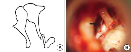
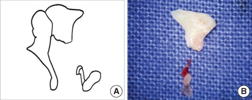
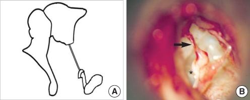
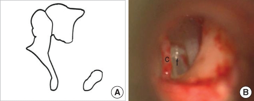
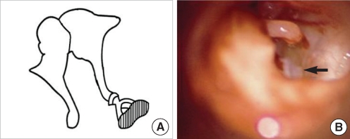
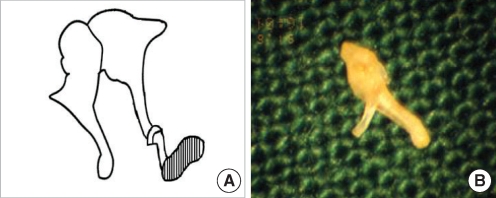
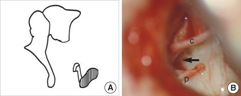
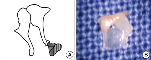
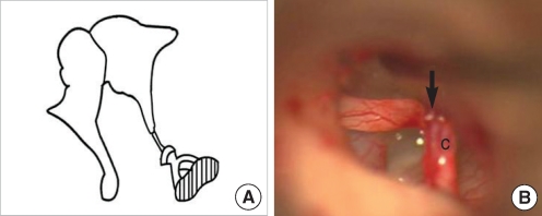
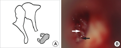
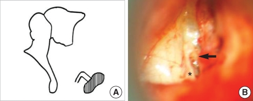
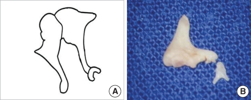
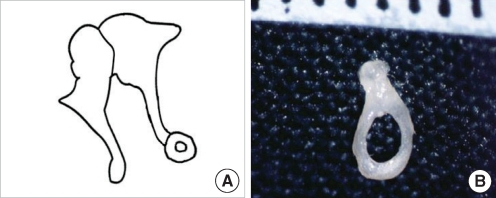
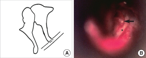
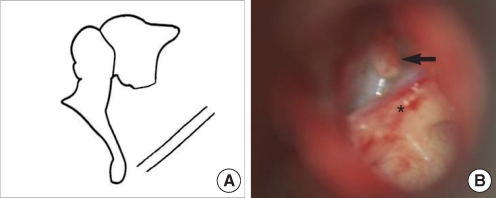
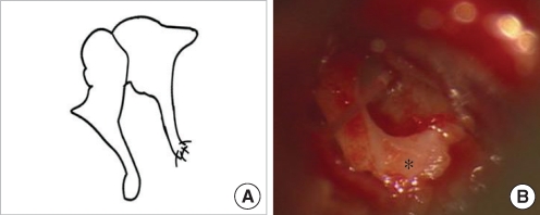
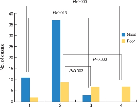
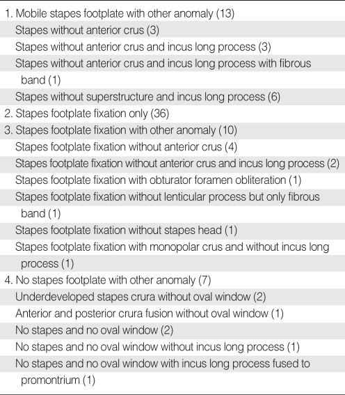
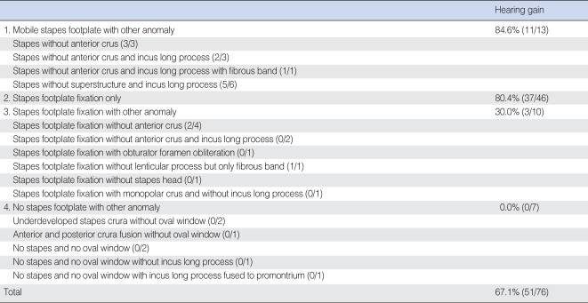
Anomalies of 66 cases could be categorized into the following 16 patterns: stapes without anterior crus (3 cases, Fig. 1); stapes without anterior crus and incus long process (3 cases, Fig. 2); stapes without anterior crus and incus long process with fibrous band (1 case, Fig. 3); stapes without superstructure and incus long process (6 cases, Fig. 4); stapes footplate fixation (36 cases, Fig. 5); stapes footplate fixation without anterior crus (4 cases, Fig. 6); stapes footplate fixation without anterior crus and incus long process (2 cases, Fig. 7); stapes footplate fixation with obturator foramen obliteration (1 case, Fig. 8); stapes footplate fixation without lenticular process but only fibrous band (1 case, Fig. 9); stapes footplate fixation without stapes head (1 case, Fig. 10); stapes footplate fixation with monopolar crus and without incus long process (1 case, Fig. 11); underdeveloped stapes crura without oval window (2 cases, Fig. 12); anterior and posterior crura fusion without oval window (1 case, Fig. 13); no stapes and no oval window (2 cases, Fig. 14); no stapes and no oval window without incus long process (1 case, Fig. 15); and no stapes and no oval window with incus long process fused to promontrium (1 case, Fig. 16). Among the above described 16 patterns of congenital stapes anomalies, stapes footplate fixation was the most common. These 16 patterns of congenital stapes anomaly were classified into 4 types according to the status of stapes footplate (Table 1). As for the stapes anomaly, 36 cases (54.5%) were bilateral (Table 2). When anomalies were confined to stapes, they were usually bilateral. In cases with only stapes footplate fixation, 31 out of 36 patients (86.1%) were bilateral. When stapes footplate fixation was combined with other anomaly, 10 out of 10 cases (100.0%) were unilateral (Table 2). Exploratory tympanotomy was performed on 76 ears of 66 patients, and hearing results according to 4 types of the status of the stapes footplate are summarized in Table 3. Mean postoperative air conduction was 29.7±15.3 dB and postoperative ABG (postoperative air conduction-preoperative bone conduction) was 13.3±13.1 dB. There was one case of postoperative sensorineural hearing loss defined as more than 15 dB loss of bone conduction after surgery. Patients with mobile stapes or only stapes footplate fixation had significantly better hearing result than patients with stapes fixation associated with other ossicular anomaly or no stapes footplate (Fig. 17). Successful hearing gain was achieved in 51 out of 76 ears (67.1%) after surgical treatment.

DISCUSSION

The upper ossicular chain is closely related more to the first branchial arch, whereas the lower ossicular chain is related to the second arch. For this reason, it is common to find malformations of the lower half of the ossicular chain in conjunction with marked stapes anomalies. Anatomic variations of the stapes may differ considerably in type and extent (11), and it is very difficult to appreciate why anomalies were involved more in the second arch-originated ossicles, restricted only to the stapes, and not involved in the other ossicles even from the same arch. Although congenital anomalies of the stapes without malformations of the external ear are not common, high resolution CT of the temporal bone and exploratory tympanotomy provide otologic surgeons with a greater chance of revealing them.
The most common congenital isolated ossicular anomalies are stapes fixation and incudostapedial discontinuity (12), and isolated congenital stapes fixation represents 20% to 35% of ossicular malformations (5, 13, 14). It is of a great interest to note that isolated stapes footplate fixation comprised more than 50% in this study. Congenital stapes fixation is the result of a fixation between the peripheral lamina stapedialis and the annular ligament (15), and it is the cause of conductive deafness with a normal tympanic membrane. The differential diagnosis of stapes fixation includes other ossicular malformations, oval or round window atresia, congenital cholesteatoma at early stage or other middle ear tumors, and ossicular trauma (7). Congenital stapes fixation must be distinguished from stapes otosclerosis which is defined by a progressive deafness, even if the surgical procedures are the same. Thus, the nonprogressive deafness is an essential element of the diagnosis, which is easy to establish, when patients are followed regularly with clinical examination and unchanged audiometric thresholds.
In this study, bilateral stapes anomaly was found in 36 out of 66 cases (54.5%), and 31 out of 36 (86.1%) cases of stapes fixation were bilateral in consistence with other studies which range from 68% to 90% (5, 7).
Another interesting finding in this study was that there were no stapes anomalies combined with the malleolar anomalies, and stapes anomalies associated with other ossicular anomalies were unilateral.
Recently, there have been increasing number of reports on the cases with stapes anomalies other than footplate fixation (16-21). However, most of stapes anomalies in these reports were found to be associated with stapes fixation. In our present study, however, 19.7% of all stapes anomaly cases involved mobile stapes footplate.
Earlier studies showed that audiometric results after surgical treatment of middle ear malformation are better for congenital stapes ankylosis, particularly if no other middle ear malformation is associated (5, 17). In this study, patients with mobile stapes or only stapes footplate fixation had significantly better hearing outcomes than patients with stapes fixation associated with other ossicular anomaly or no stapes footplate. Furthermore, Teunissen et al. (5) reported poor surgical outcome in cases associated with stapes superstructure anomaly, in agreement with our result. Therefore, it is quite likely that well developed stapes is important in hearing improvement.

CONCLUSION

In this study, we found that most of the footplate fixation were bilateral, whereas stapes anomalies associated with other ossicular anomaly were unilateral. The success of the surgical treatment of stapes anomaly might depend on developmental status of the footplate. Stapes anomalies were detected with no fixed patterns, therefore, it is expected to detect a large variety of these patterns in future.

CONFLICT OF INTEREST

All authors had no conflict of interest on this study.

References

1. House HP, House WF, Hildyard VH. Congenital stapes footplate fixation: a preliminary report of twenty-three operated cases. Laryngoscope. 1958 8;68(8):1389-1402. PMID: 13576898.
crossref pmid
2. Lindsay JR, Sanders SH, Nager GT. Histopathologic observations in so-called congenital fixation of the stapedial footplate. Laryngoscope. 1960 12;70(12):1587-1602. PMID: 13762396.
crossref pmid
3. Gundersen T. Congenital malformations of the stapes footplate. Arch Otolaryngol. 1967 2;85(2):171-176. PMID: 6017589.
crossref pmid
4. Steele BC. Congenital fixation of the stapes footplate. Acta Otolaryngol. 1969;(Suppl 245):1-24. PMID: 5379836.

5. Teunissen B, Cremers WR, Huygen PL, Pouwels TP. Isolated congenital stapes ankylosis: surgical results in 32 ears and a review of the literature. Laryngoscope. 1990 12;100(12):1331-1336. PMID: 2243529.
crossref pmid
6. Nandapalan V, Tos M. Isolated congenital stapes ankylosis: an embryologic survey and literature review. Am J Otol. 2000 1;21(1):71-80. PMID: 10651438.
crossref pmid
7. Albert S, Roger G, Rouillon I, Chauvin P, Denoyelle F, Derbez R, et al. Congenital stapes ankylosis: study of 28 cases and surgical results. Laryngoscope. 2006 7;116(7):1153-1157. PMID: 16826051.
crossref pmid
8. Schlosser WD, Goldman BR, Winchester RA. Further experiences with the diagnosis and surgical management of congenital mixed deafness. Laryngoscope. 1964 6;74:773-789. PMID: 14171385.
crossref pmid
9. Hashimoto S, Yamamoto Y, Satoh H, Takahashi S. Surgical treatment of 52 cases of auditory ossicular malformations. Auris Nasus Larynx. 2002 1;29(1):15-18. PMID: 11772485.
crossref pmid
10. Committee on Hearing and Equilibrium. Committee on hearing and equilibrium guidelines for the evaluation of results of treatment of conductive hearing loss. Otolaryngol Head Neck Surg. 1995 9;113(3):186-187. PMID: 7675477.
crossref pmid pdf
11. Dass R, Grewal BS, Thapar SP. Human stapes and its variations. II. Footplate. J Laryngol Otol. 1966 5;80(5):471-480. PMID: 5934111.
crossref pmid
12. Yuen HY, Ahuja AT, Wong KT, Yue V, van Hasselt AC. Computed tomography of common congenital lesions of the temporal bone. Clin Radiol. 2003 9;58(9):687-693. PMID: 12943639.
crossref pmid
13. Raveh E, Hu W, Papsin BC, Forte V. Congenital conductive hearing loss. J Laryngol Otol. 2002 2;116(2):92-96. PMID: 11827579.
crossref pmid
14. Hung KL, Tu TY, Tsai TL, Shiao AS. Congenital ossicular anomalies. J Chin Med Assoc. 2003 8;66(8):474-479. PMID: 14604311.
pmid
15. Dornhoffer JL, Helms J, Hoehmann DH. Stapedectomy for congenital fixation of the stapes. Am J Otol. 1995 5;16(3):382-386. PMID: 8588635.
pmid
16. Cremers CW, Hoogland GA. Congenital stapes ankylosis by elongation of the pyramidal eminence. Ann Otol Rhinol Laryngol. 1986;Mar–Apr;95(2 Pt 1):167-168. PMID: 3963687.
crossref pmid
17. Teunissen B, Cremers CW. Surgery for congenital stapes ankylosis with an associated congenital ossicular chain anomaly. Int J Pediatr Otorhinolaryngol. 1991 5;21(3):217-226. PMID: 1869375.
crossref pmid
18. Kinsella JB, Kerr AG. Familial stapes superstructure fixation. J Laryngol Otol. 1993 1;107(1):36-38. PMID: 8445310.
crossref pmid
19. Nandapalan V, Tos M. Isolated congenital stapes suprastructure fixation. J Laryngol Otol. 1999 9;113(9):798-802. PMID: 10664680.
crossref pmid
20. Doi T, Nakazawa H, Adachi M, Kaneko T, Munemoto Y, Komeda M, et al. Familial isolated stapes-pyramidal fixation by a bony bar with normal stapedius tendon. Arch Otolaryngol Head Neck Surg. 2005 4;131(4):349-352. PMID: 15837906.
crossref pmid
21. Ching HY, Bottrill ID. An unusual stapes. Surg Radiol Anat. 2006 10;28(5):474-476. PMID: 16826320.
crossref pmid
Fig. 1
Stapes without anterior crus. (A) Schematic presentation. (B) Operative finding shows no anterior crus of the stapes (arrow).
c: chorda tympani.
ceo-2-33-g001.jpg
Fig. 2
Stapes without anterior crus and incus long process. (A) Schematic presentation. (B) Removed incus shows no long process, and removed stapes shows no anterior crus.
ceo-2-33-g002.jpg
Fig. 3
Stapes without anterior crus and incus long process with fibrous band. (A) Schematic presentation. (B) Operative finding shows a fibrous band (arrow) instead of the incus long process and the stapes lacking anterior crus (asterisk).
ceo-2-33-g003.jpg
Fig. 4
Stapes without superstructure and incus long process. (A) Schematic presentation. (B) Operative finding shows no incus long process and no stapes superstructure.
c: chorda tympani; f: footplate.
ceo-2-33-g004.jpg
Fig. 5
Stapes footplate fixation. (A) Schematic presentation. (B) Operative finding shows half of the stapes footplate remaining (arrow) because of partial fixation of its footplate.
ceo-2-33-g005.jpg
Fig. 6
Stapes footplate fixation without anterior crus. (A) Schematic presentation. (B) Removed stapes shows an underdeveloped anterior crus.
ceo-2-33-g006.jpg
Fig. 7
Stapes footplate fixation without anterior crus and incus long process. (A) Schematic presentation. (B) Operative finding shows no incus long process and the stapes lacking anterior crus (arrow) without mobility of its footplate. c: chorda tympani; p: posterior crus.
ceo-2-33-g007.jpg
Fig. 8
Stapes footplate fixation with obturator foramen obliteration. (A) Schematic presentation. (B) Removed stapes shows a non-perforated obturator foramen.
ceo-2-33-g008.jpg
Fig. 9
Stapes footplate fixation without lenticular process: only fibrous band. (A) Schematic presentation. (B) Operative finding shows a fibrous band (arrow) instead of the lenticular process of the incus.
c: chorda tympani.
ceo-2-33-g009.jpg
Fig. 10
Stapes footplate fixation without stapes head. (A) Schematic presentation. (B) Operative finding shows only anterior (white arrow) and posterior crus (black arrow) without mobility of its footplate.
ceo-2-33-g010.jpg
Fig. 11
Stapes footplate fixation with monopolar crus and without incus long process. (A) Schematic presentation. (B) Operative finding shows only one crus in the center of the footplate (arrow) and stapes-pyramidal fixation by a bony bar (asterisk).
ceo-2-33-g011.jpg
Fig. 12
Underdeveloped stapes crura without oval window. (A) Schematic presentation. (B) Removed stapes shows underdeveloped crura.
ceo-2-33-g012.jpg
Fig. 13
Anterior and posterior crura fusion without oval window. (A) Schematic presentation. (B) Removed stapes shows a crural fusion.
ceo-2-33-g013.jpg
Fig. 14
No stapes and no oval window. (A) Schematic presentation. (B) Operative finding shows a long process of the incus (arrow) without the stapes, which is in contact with a downward facial nerve (asterisk) in the mesotympanum.
ceo-2-33-g014.jpg
Fig. 15
No stapes and no oval window without incus long process. (A) Schematic presentation. (B) Operative finding shows shortening of the incus long process (arrow) without the stapes and downward displacement of the tympanic portion of the facial nerve with a bony bar (asterisk).
ceo-2-33-g015.jpg
Fig. 16
No stapes and no oval window with incus long process fused to promontorium. (A) Schematic presentation. (B) Operative finding shows the long process of the incus fused to promontorium (asterisk) without the stapes and the oval window.
ceo-2-33-g016.jpg
Fig. 17
Comparison of postoperative hearing results of congenital stapes anomaly according to status of stapes footplate (76 ears). Patients with mobile stapes or only stapes footplate fixation had significantly better hearing result than patients with stapes fixation associated with other ossicular anomaly or no stapes footplate.
1: Mobile stapes footplate with other anomaly; 2: Stapes footplate fixation only; 3: Stapes footplate fixation with other anomaly; 4: No stapes footplate with other anomaly; Good: postoperative air-bone gap <20 dB; Poor: postoperative air-bone gap ≥20 dB. Postoperative air-bone gap: postoperative air conduction-preoperative bone conduction.
ceo-2-33-g017.jpg
Table 1
Patterns of congenital stapes anomaly according to status of stapes footplate (66 cases)
ceo-2-33-i001.jpg
Table 2
Bilaterality of stapes anomaly according to status of stapes footplate
ceo-2-33-i002.jpg
Table 3
Hearing results of congenital stapes anomaly according to status of stapes footplate after more than 3 months of surgery (76 ears)
ceo-2-33-i003.jpg

Hearing gain; postoperative air conduction-preoperative bone conduction <20 dB.

TOOLS
Download PDF File  Download PDF File
PubReader  PubReader
ePub Link  ePub Link
XML Download  XML Download
Full text via DOI  Full text via DOI
Download Citation  Download Citation
  Print
Share:      
METRICS
28
Crossref
29
Scopus
10,723
View
138
Download
Related article
Editorial Office
Korean Society of Otorhinolaryngology-Head and Neck Surgery
103-307 Park Tower officetel, Yongsan-dong 5-ga, Yongsan-gu, Seoul 04385, Korea
TEL: +82-2-711-9091   FAX: +82-2-3487-6603   E-mail: editor.eceo@gmail.com
Copyright © Korean Society of Otorhinolaryngology-Head and Neck Surgery.                 Developed in M2PI
Close layer
prev next