INTRODUCTION
Rhinoplasty is considered to be a popular and effective form of cosmetic surgery [1]. Nevertheless, its postoperative complications may include significant eyelid edema and ecchymosis [2]. Periorbital edema may cause difficulties in visual acuity in the early postoperative period, and ecchymosis may increase periorbital pigmentation and cause disruptions in the patientŌĆÖs social activities. The main cause of eyelid edema and ecchymosis after rhinoplasty is soft tissue damage at the osteotomy site [2]. Careful and delicate surgical manipulation can help to reduce such damage, but it cannot be completely prevented.
External percutaneous osteotomy and the internal endonasal technique are the two most common approaches used in rhinoplasty. Considering the risk of periosteal injury and the manipulation of the surrounding soft tissue during osteotomy, lateral osteotomy can be done with or without periosteal elevation (subperiosteal tunneling) [3]. However, there is no consensus regarding which osteotomy method is more effective for reducing morbidity in rhinoplasty procedures. Since rhinoplasty remains a challenging procedure to this day, it is important for clinicians to follow the best practices for reducing postoperative complications. This review aimed to evaluate the efficacy of periosteal elevation. To assess the evidence for periosteal elevation prior to lateral osteotomy, we conducted a review of the literature to identify studies investigating eyelid edema, ecchymosis, and subconjunctival hemorrhage in patients undergoing lateral osteotomy with or without periosteal elevation.
MATERIALS AND METHODS
Search strategy and selection of studies
Studies published in English until May 2019 from Medline, Scopus, and Cochrane Register of Controlled Trials were identified. Search terms were ŌĆ£rhinoplasty,ŌĆØ ŌĆ£osteotomy,ŌĆØ ŌĆ£periosteum,ŌĆØ ŌĆśedema,ŌĆØ and ŌĆ£ecchymosis.ŌĆØ We also checked the reference lists of identified studies to ensure that relevant studies were not missed.
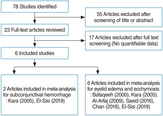
Two reviewers (JSK, SHH) independently screened all abstracts and titles for candidate studies, excluding those that were not associated with lateral osteotomy of rhinoplasty. Randomized controlled trials that studied patients undergoing rhinoplasty and periosteal elevation or periosteal preservation were included. Studies were excluded from analysis if patients underwent additional surgery (such as blepharoplasty or orthognathic surgery) or if reports were duplicated or if the results were not clearly reported as quantifiable data, or if appropriate data could not be extracted and calculated from published results. Fig. 1 shows the search strategy for meta-analysis.
Data extraction and risk of bias assessment
Data from included studies were extracted using standardized forms. Primary outcomes were degree of eyelid edema [3-6], ecchymosis [3-8], and subconjunctival hemorrhage [3,4] comparing different osteotomy methods with another methods during the postoperative period (within 3 days or the seventh postoperative day). Eyelid edema and ecchymosis were assessed individually using visual grading scales. On the grading scale, the smallest number indicated no edema or ecchymosis, and the largest number indicated severe edema enough to close the eyelid or severe ecchymosis spreading to the lateral canthus.
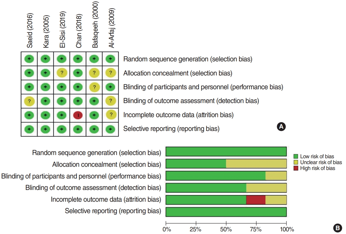
In studies of the effect of subperiosteal tunneling on measured outcomes, data on the number of patients, grading scale, and P-value were extracted. Quality assessment of the included randomized controlled studies was performed using the Cochrane risk of bias tool. We used a ŌĆ£risk of biasŌĆØ table including random sequence generation, allocation concealment, blinding, incomplete outcome data, and free of selective reporting (Fig. 2).
Statistical analysis
A meta-analysis of the selected studies was conducted by R statistical software (R Foundation, Vienna, Austria) with continuous measurements comparing the mean and standard deviation between the control and experimental groups. The effect sizes of edema, ecchymosis, and subconjunctival hemorrhage were expressed as the standardized mean difference (SMD). Heterogeneity was confirmed by Cochran Q statistic test and I2 test. In this analysis, outcomes that did not show heterogeneity (I2<50) were analyzed using fixed-effects model.
RESULTS
Six studies with a total of 208 patients were included in this meta-analysis. Due to the incomplete reporting of patient variables in these studies, a clear comparison of patientsŌĆÖ overall characteristics could not be made. The characteristics of the included studies are shown in Table 1.
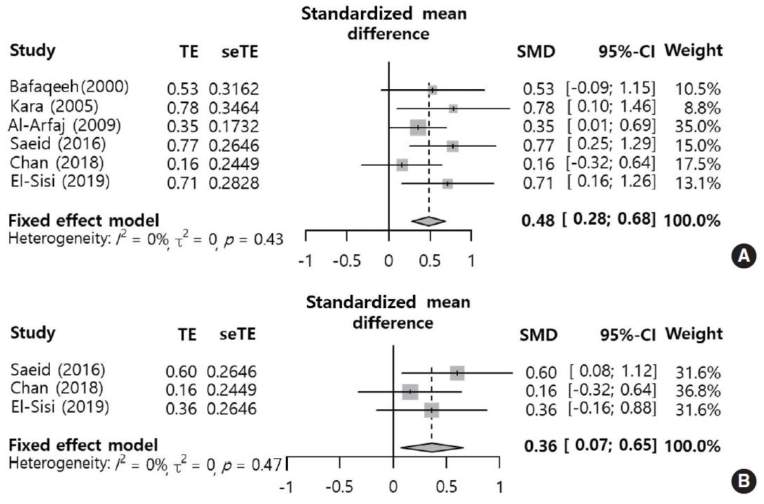
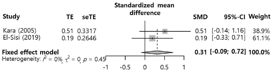
Eyelid edema (SMD, 0.45; 95% confidence interval [CI], 0.18 to 0.72; I2=35%) within 3 days postoperatively was significantly more common in the periosteal elevation group than in the preservation group. However, edema on postoperative day 7 (SMD, 0.21; 95% CI, ŌĆō0.09 to 0.50; I2=0%) showed no significant difference between the two groups (Fig. 3). Eyelid ecchymosis was significantly more common in the periosteal elevation group than in the preservation group (within 3 days postoperatively: SMD, 0.48; 95% CI, 0.28 to 0.68; I2=0%; on postoperative day 7: SMD, 0.36; 95% CI, 0.07 to 0.65; I2=0%) (Fig. 4). There was no significant difference in subconjunctival hemorrhage on day 1 (SMD, 0.31; 95% CI, ŌĆō0.09 to 0.72; I2=0%) (Fig. 5). No significant inter-study heterogeneity was found for these outcomes (I2<50%).
DISCUSSION
Repositioning or changing the location of the nasal bone is essential in most rhinoplasty procedures performed for external correction [9]. In particular, medial movement of the nasal bone via osteotomy allows the pyramid to be reconstructed to its normal condition [10]. Osteotomy is performed with the goals of straightening a deviated dorsum, narrowing the nasal dorsum, and opening the nasal vault [11]. Eyelid edema and ecchymosis, which are the most common postoperative complications of osteotomy, can be caused by damage to arteries or veins and soft tissue injuries during lateral osteotomy [5]. In lateral osteotomy, considerable force is delivered to the soft tissues and bones to mobilize the nasal bone. Thus, lateral osteotomy should be performed safely and accurately with minimal postoperative complications while producing aesthetically satisfactory results [12,13]. In order to minimize the complications of lateral osteotomy, various innovations have been made in surgical instruments, preoperative local infiltration, and intraoperative techniques [7,14,15].
Lateral osteotomy can be performed after periosteal elevation. A subperiosteal tunnel is formed by elevating the periosteum and upper layer from the bony cortex in the path of the osteotomy [5]. The goal of subperiosteal tunneling is to protect the blood vessels and to reduce periosteal injury during osteotomy [16]. This technique uses an elevator to make the subperiosteal tunnel wide enough for the osteotome to enter. This prevents the osteotome from damaging the soft tissue, resulting in a bloodless osteotomy [15,17,18]. The elevated periosteum also becomes a barrier that prevents blood from spreading into subcutaneous tissues [16]. However, several studies have suggested that periosteal elevation is more likely to cause damage to vessels [3,4,7]. In addition, subperiosteal tunneling can damage the lacrimal sac and the canthal ligament because the medial canthal ligament is just above the lacrimal sac [10,12]. Therefore, elevation of the periosteum may be unsafe because it can damage surrounding structures.
Edema and ecchymosis have been reported to reach maximum severity within 3 days postoperatively although they may persist until the 9th postoperative day [19]. Therefore, we used two time points to investigate changes in these outcomes over time. Periosteal elevation was found to be associated with more frequent edema and ecchymosis (postoperative edema within 3 days and postoperative ecchymosis at both time points), except for eyelid edema on postoperative day 7. In particular, the SMDs of the edema and ecchymosis outcomes within 3 days after surgery were near 0.5, with clinically medium effect sizes [20]. Because periosteal elevation requires more manipulation of the tissue, it may increase the risk of periorbital edema and ecchymosis [3]. Subperiosteal tunnels can also serve as a potential space for blood to accumulate and spread into the surrounding tissue [3,7]. Although periosteal elevation is designed to protect blood vessels, it can simultaneously provide a space for hematoma to spread more widely. Some studies have suggested that complications may be more frequent if the stabilization and functioning of the periosteum are completely stopped by elevation, rather than partial damage [21,22]. These possible explanations provide support for our results on postoperative edema and ecchymosis.
Periorbital ecchymosis is not limited to the subcutaneous tissue of the eyelids; instead, it sometimes continues through the orbital adipose tissue, resulting in subconjunctival ecchymosis [23]. Considering the positive effect of periosteal preservation on eyelid edema and ecchymosis, subconjunctival ecchymosis would be expected to be significantly less frequent in the periosteal preservation group than in the periosteal elevation group. In contrast to our expectations, postoperative ecchymosis on day 1 showed no significant difference between the two groups. However, this result was only based on two studies. Thus, the clinical implications of this result may not be meaningful, which is a limitation of our findings.
A limitation of our study is that surgical manipulations other than subperiosteal tunneling might also increase postoperative complications. In addition, when blood accumulates in surrounding tissues as a result of extensive bleeding during surgery, periorbital ecchymosis may be more likely. The results of our analysis could not distinguish these factors affecting complications that are distinct from subperiosteal tunneling. Nonetheless, in each of the studies that we included, there was no difference in the management of the two groups, enabling a meaningful comparison of the outcome measures. We considered that using the SMD to compare two groups under the same conditions was adequate from a methodological perspective. The spread of edema or bleeding could also vary depending on individual-level variations in skin thickness and the amount of subcutaneous fat [4,24]. If further studies are conducted on this issue, it may be possible to obtain more reliable results.
HIGHLIGHTS
Ō¢¬ Eyelid edema within 3 days after rhinoplasty was significantly more common in the periosteal elevation group than in the periosteal preservation group.
Ō¢¬ Eyelid ecchymosis was significantly more common in the periosteal elevation group than in the periosteal preservation group, both within 3 days and at 7 days after rhinoplasty.
Ō¢¬ Periosteal preservation during osteotomy may reduce eyelid edema and ecchymosis compared to periosteal elevation.