Externally Monitored Versus Conventional Buried Flaps in Laryngopharyngeal Reconstruction
Article information

Abstract
Objectives
To compare the surgical outcomes of externally monitored and conventional buried flaps with the goal of determining the usefulness of external monitoring of buried flaps.
Methods
In this case-control study with propensity score matching, 30 patients were evenly divided into externally monitored buried flap and conventional buried flap groups. The total operative time for free flap reconstruction, the flap survival rate, the length of hospital stay, the initial time of a reliable visual assessment, complications, the final diet achieved, and the duration until diet initiation were compared between the groups.
Results
The mean operative time for reconstruction was 115 minutes (interquartile range, 85–150 minutes) and 142 minutes (interquartile range, 95–180 minutes) in the externally monitored and conventional groups, respectively (P= 0.245). The median length of hospital stay was 24 days (interquartile range, 18–30 days) and 27 days (interquartile range, 20–41 days) in the externally monitored and conventional groups, respectively (P=0.298). The median duration until diet initiation was 15 days (interquartile range, 15–21 days) and 18 days (interquartile range, 15–34 days) in the externally monitored and conventional groups, respectively (P=0.466). The final diet, initial time of a reliable visual assessment, and complications were comparable between the groups, but the external skin paddle provided an excellent visual assessment immediately postoperatively in all cases.
Conclusion
The outcomes were comparable between the groups, indicating that externalization of the cutaneous component of a buried flap may be a straightforward and useful technique for monitoring a buried anterolateral thigh free flap in laryngopharyngeal reconstructions. The salvage and false-positive rates of compromised flaps should be compared in large subject groups in future studies to prove that the use of an external skin paddle improves flap monitoring.
INTRODUCTION
Total laryngectomy with partial or total pharyngectomy is the mainstay treatment for advanced laryngeal and hypopharyngeal squamous cell carcinoma. In general, laryngopharyngeal defects should be primarily reconstructed to provide alimentary tract continuity and to restore speech and swallowing functions. Microvascular free tissue transfer is currently the preferred reconstruction method for such defects. With recent surgical and technical developments in microsurgical flap procedures, the success rates for these procedures are estimated to be over 95%; however, there is still a possibility of failure [1]. Flap failure is attributed to arterial or venous occlusion arising from thrombosis, external compression, vessel kinking, or hematoma. Microvascular thrombosis occurs in 4% of flaps, and in such cases, the earliest possible revision of microanastomosis provides the best chance for flap salvage [2].
Although reconstructive surgeons have used a broad range of technologies to monitor pedicle and flap perfusion continuously, clinical assessments such as monitoring skin color, turgor, temperature of the flap, and capillary refill have yielded the best outcomes [1]. Disa et al. [3] reported that regular monitoring during the first few days postoperatively yielded flap salvage rates of up to 80% and overall success rates of up to 99% [4].
In laryngopharyngeal reconstructions, a free flap necessitates a buried flap, which precludes clinical monitoring. The inability to monitor buried free flaps delays the early identification of vascular compromise, which generally occurs within 48 hours. Thus, the flap salvage rate and flap loss rate of buried flaps are significantly higher than those of non-buried free flaps [3]. Externalization of a segment of the skin paddle from the buried free flap is known to improve clinical monitoring of the buried free flap. Considering this, various trials have aimed to externalize a portion of the free flap [5,6]. Although some studies have shown a higher fistula rate for pharyngeal reconstructions using a monitored skin paddle [7], we experienced several successful consecutive cases in which a deepithelialized bridge was used to externally monitor a part of the anterolateral thigh free flap. To our knowledge, no matched comparative study on externally monitoring a buried flap of the anterolateral thigh free flap has been performed. Thus, the aim of this study was to compare the clinical outcomes of an externally monitored buried flap to those of a conventional buried anterolateral thigh free flap in laryngopharyngeal reconstructions performed by a single experienced surgeon through a matched case-control study with propensity score matching. The use of a propensity score is appropriate for a retrospective comparative study, as it reduces the risk of confounding factors between different groups.
MATERIALS AND METHODS
Ethical considerations
This retrospective comparative analysis was conducted with the approval of the Institutional Review Board of Severance Hospital (IRB No. 4-2020-0750) and complied with the ethical principles of the Declaration of Helsinki for medical research involving human subjects. All patients provided written informed consent.
Patients
The medical records of 50 patients who underwent laryngopharyngeal reconstruction with an anterolateral thigh free flap by a single surgeon (WSK) in Severance Hospital (Seoul, Korea) were retrospectively reviewed without exclusion. The patients were classified according to whether they had the conventional buried flap or external monitored flap including an externally placed cutaneous monitoring paddle because of advanced head and neck cancers between July 2013 and February 2017. From October 2015, the surgeon started to use an externally monitored buried flap in consecutive cases, with some exceptions decided on a case-by-case basis. Altogether, there were 46 men and four women. Fifty laryngopharyngeal defects were analyzed with respect to sex, age, etiology, operative time, length of hospitalization, timing of oral diet initiation, highest level of diet achieved postoperatively, and complications.
Surgical technique
In the case of an external monitored buried flap, the donor site is harvested as per the modified design on the anterolateral thigh based on the anticipated perforator location, and it includes a deepithelialized bridge and a monitoring flap measuring 2 cm (Fig. 1). After dissecting along the perforator, the skin paddle is manipulated to construct a deepithelialized bridge between the neopharynx and monitoring flap to facilitate sharing of a perforator (Fig. 2). The free flap is transferred to the defect, skin of the anterolateral thigh free flap is placed so it faces the pharyngeal lumen, bridge is folded once, and free margin of the monitoring flap is placed so it faces the tracheostoma (Fig. 3). To ensure good pedicle geometry and adequate pedicle length while minimizing post-anastomotic manipulation, insetting of the inferior and lateral margins of the neopharynx is prioritized over microanastomosis. The complete insetting of the free flap is performed after microanastomosis with reinforcement using the fascial layer. The upper neck skin flap is redraped over the neopharynx, and the monitoring segment is externalized through the neck incision and sutured superiorly to the permanent tracheostoma. While preserving the larynx, the monitoring flap is brought out through the neck incision and exposed externally (Fig. 4). In other cases, the conventional buried flap is used as a classic technique with no externalized part [8].

Preoperative design of an externally monitored buried flap on the anterolateral thigh.
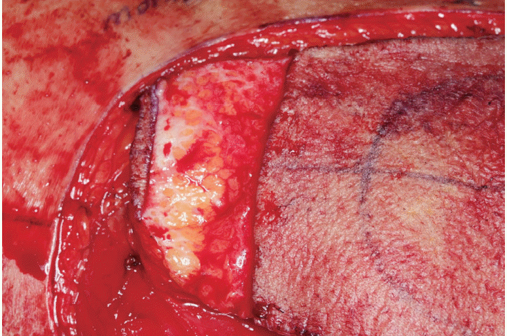
Harvested free anterolateral thigh flap after making a de-epithelialized bridge.

Postoperative externalized viable skin paddle sutured on the upper side of the tracheostoma after total laryngectomy with partial pharyngectomy.

Healed externally monitored buried flap. (A) Upper side of the tracheostoma after total laryngectomy with partial pharyngectomy. (B) Gap between the transverse skin incision after partial laryngectomy without pharyngectomy.
Postoperative follow-up
Most patients were kept sedated on a ventilator overnight in a surgical intensive care unit and weaned off the ventilator support the next day. Typically, the free flaps were monitored hourly for the first 24 hours, with tapering of the frequency thereafter, to identify vascular compromise as soon as possible so that salvage action could be taken. The externalized segment was monitored by clinical assessment of the skin color, turgor, temperature of the flap, skin prick test result (Fig. 5), and capillary refill, but the conventional buried flap was monitored visually by a fiberoptic laryngoscope because of poor accessibility. Through the endoscopic or naked-eye view, the surgeon recorded the initial time of reliable free flap assessment postoperatively. Acoustic Doppler ultrasonography, covered skin flap, and drained fluid status were referred to in the evaluation of a compromised flap. A compromised flap is defined as a postoperative complication requiring unplanned repeat surgery to salvage the unhealthy flap or avoid flap loss. Tube feeding was started on postoperative day 1 and continued until clinical swallow evaluation, which was generally performed on postoperative day 14. When a radiographic leak or fistula was identified, patients continued feeding tube therapy for an additional week, and an evaluation was repeated until the leak had healed. Swallowing function was assessed by the type of diet, which was categorized as a general diet (solid food), soft diet, liquid diet, or as being partially or totally tube-feeding dependent.

Positive pin-prick test of the externally monitored buried flap on the third postoperative day.
Statistical analysis
To perform this matched case-control study with reduced selection bias, we used a logistic regression model to estimate the propensity score and included variables that can affect the outcome of surgery (sex, age, body mass index, T stage, and flap size) and matched the patients in 1:1 ratio based on a caliper of 0.1% [9]. Twenty patients were excluded in the conventional buried flap population to match propensity score. Post-matching data were analyzed with Fisher’s exact test, the independent t-test, and MannWhitney U-test. The relative risk was calculated to compare complications between the groups. All tests were two-sided, with a level of significance set at P<0.05. Statistical analyses were performed by an expert in biomedical statistical analysis who used IBM SPSS ver. 21.0 (IBM Corp., Armonk, NY, USA).
RESULTS
All the externally monitored buried flaps were monitored carefully with clinical assessments, especially the skin prick test. The conventional buried flap group and the externally monitored buried flap group were compared with respect to the characteristics of each set of 15 patients with a matched propensity score who underwent total laryngectomy with partial pharyngectomy, partial laryngectomy without pharyngectomy, or partial pharyngectomy without laryngectomy (Table 1). The mean age of the patients in the conventional buried flap and external monitored buried flap groups was 62.7±7.2 and 63.8±8.0 years, respectively. Hypopharyngeal cancer was the most common primary lesion in both groups. The flaps in the externally monitored buried flap group were larger (66.20±27.63 cm2) than those in the conventional buried flap group (59.58±24.87 cm2) because of the external skin paddle; however, the difference was not statistically significant. There were no statistically significant differences with respect to the total reconstruction time, duration of hospitalization, and mean timing of oral diet initiation between the two groups. A general diet was the most common type of diet, with no significant difference between the groups. Complications such as flap compromise, pharyngocutaneous fistula, stomal and pharyngeal stenoses, and bleeding were comparable between the two groups. The relative risk of all complications was either the same or less in the external monitored buried flap group. Although a reliable endoscopic visual assessment was difficult in both groups initially, the external skin paddle provided excellent visual assessment immediately postoperatively in all cases (Table 2).

Clinicopathologic characteristics of the patients

Perioperative parameters and postoperative complications, with the calculated relative risk for an externally monitored buried flap
DISCUSSION
Synopsis of key findings
Our findings suggest that there are no evident differences in surgical outcomes between external monitored buried flaps and conventional buried flaps in terms of the operative time, flap failure, fistula rate, and swallowing function. An external monitored buried flap provided reliable flap monitoring without false results, such as a viable flap with a non-viable external monitoring part.
Strengths of the study
Because of differences in surgeons’ surgical skills and variety in patients’ lesions, there have been few comparative studies of external monitored buried flaps and conventional buried flaps. The strengths of the present study include that the procedures were performed by a single surgeon and propensity score matching was conducted using factors (e.g., sex, age, body mass index, T stage, and flap size) that could potentially compromise free flaps; thereby, we were able to reduce selection bias. Another strength of our study is that by comparing the reliable visual assessment time, we were able to clearly identify the advantages of flap monitoring in the external monitored buried flap group. Finally, this study indicates that there is no need for additional equipment, such as an implantable Doppler system for buried flaps, because external monitoring is a cost-effective, straightforward, and useful way to monitor buried flaps.
Comparisons with other studies
Lindau et al. [10] reported the largest series on the use of buried free flaps (n=103) in the head and neck regions. They concluded that buried flaps did not have a higher failure rate than other flaps and that an implantable Doppler system was helpful for monitoring buried free flaps. According to their study, no further external monitoring is required; however, their study was limited in evaluating the risk of laryngopharyngeal defects in buried flaps because only 34 laryngopharyngeal defects were evaluated using heterogeneous free flaps, without a control group. Moreover, considering that most salvage operations (3/4, 75%) were performed to salvage compromised flaps of laryngopharyngeal defects, buried flaps for laryngopharyngeal defects are more likely to be compromised, higher possibility to be failed than other buried flaps. Contrary to the results of Lindau et al. [10], Disa et al. [3] reported a higher failure rate of buried flaps (n=77, including flaps in the head and neck, trunk and breast, and extremities) due to late re-exploration, usually >7 days, in patients presenting with a wound infection or fistula. Given this controversy about the failure rate and the known fact that laryngopharyngeal defects are likely to cause life-threatening fistula complications resulting from saliva [11,12], our comparative study is valuable in that we evaluated whether an external monitored buried flap affected the success of free flaps and whether it can be considered a reliable monitoring technique in laryngopharyngeal reconstruction.
The primary limitation of our study is the small number of cases of conventional buried flaps and externally monitored flaps. We present a comparison of the salvage rate and false-positive rate of compromised flaps between the two groups in Table 2, as these parameters are essential for comparing the monitoring techniques; however, a larger number of subjects would have been needed to reach statistical significance [1]. Moreover, a propensity score-matched analysis with a small sample size should be performed carefully; however, if the true confounding factor related to the outcome is included, this type of study can yield a correct estimation of the experimental treatment effect [13]. Chang et al. [14] reviewed the risk factors associated with flap loss and salvage in free flap head and neck reconstructions (n= 2,296). Age, body mass index, and comorbidities associated with patient-related risk factors did not affect flap salvage; nevertheless, for matching, we used patient-related variables (i.e., sex, age, body mass index, the T stage, and flap size) in consideration of their potential relationship with the surgical outcome. The surgical technique and experience were not assessed because the reconstructions in this study were performed by a single experienced surgeon who used the same surgical and monitoring techniques; however, we controlled for the T stage and flap size, as intraoperative confounding factors associated with the reconstruction size, in order to reduce potential selection bias. In our study, interpreting the comparative data on flap compromise was impossible because there was only one case in which the flap was compromised in the conventional buried group. Based on the knowledge of the significantly poorer outcomes of late takebacks (>3 days) of compromised flaps [15], a comparison of the takebacks of multiple salvage cases between the groups would have more clearly proven the advantages of an externally monitored buried flap. Finally, because of the lack of numbers to fill in the cross-table to evaluate the diagnostic power of externally monitored buried flaps, false-positive or false-negative values could not be calculated in this study. Moreover, monitoring of flaps can yield false-negative findings. For example, partial ischemia of the distal part may be difficult to distinguish from complications of vascular anastomosis. Therefore, a future comparative study with a large number of cases should elucidate the possibility of false-negatives when this method is used.
In conclusion, close monitoring is a key component of postoperative care following free tissue transfer. However, completely buried flaps can be difficult to observe without appropriate equipment. We suggest that externalization of the skin paddle of buried flaps is an easy and useful technique for monitoring anterolateral thigh free flaps in laryngopharyngeal reconstructions, as there were no differences in outcomes compared with conventional buried flaps and the external skin paddle provided a superior visual assessment. This external monitoring technique can be used routinely to monitor the buried flap in procedures performed to reconstruct laryngopharyngeal defects, especially for less experienced surgeons.
HIGHLIGHTS
▪ Outcomes were not different between external monitored and conventional buried flaps.
▪ Externalization of the cutaneous component of such flaps may be a straightforward and useful technique.
▪ An externally monitored buried flap allows reliable flap monitoring.
▪ Future studies should compare the salvage and false-positive rates using large samples.
Notes
No potential conflict of interest relevant to this article was reported.
Acknowledgements
This research was supported by the Basic Science Research Program through the National Research Foundation of Korea (NRF) funded by the Ministry of Education (NRF-2019R1I1A3A010-63629). This work was also supported by the Soonchunhyang University Research Fund.
Notes
AUTHOR CONTRIBUTIONS
Conceptualization: MJB, WSK. Data curation: GN. Formal analysis: MJB, GN, JK, NHH. Funding acquisition: MJB, WSK. Methodology: MJB, WSK. Project administration: ECC, WSK, JHP. Visualization: WSK. Writing–original draft: MJB. Writing–review & editing: MJB, SK, WSK.