| Home | E-Submission | Sitemap | Contact Us |  
Clinical and Experimental Otorhinolaryngology > Volume 15(4); 2022 > Article
Lee, Volpe, and Chang: The Role of Viruses in the Inception of Chronic Rhinosinusitis

Abstract

Chronic rhinosinusitis (CRS) is a complex inflammatory disorder that affects between 2% and 16% of adults in the United States, with estimated healthcare costs between 4 and 12 million USD. Viruses are a common etiologic factor for URIs, are frequently identified in the sinuses of patients with CRS, and trigger CRS exacerbations. Therefore, investigating the role of viruses provides an opportunity to identify their role in the pathogenesis of CRS. In this review, we identified the viruses frequently isolated in patients with CRS, as well as their associated immunologic responses and contributions to inflammation. Rhinovirus, parainfluenza virus, influenza virus, and respiratory syncytial virus are the viruses commonly found in patients with CRS. This information allows us to target pathways early in the pathogenesis of CRS, thereby playing a significant role in slowing the progression of this chronic disease.

INTRODUCTION

Studies on the pathogenesis of chronic rhinosinusitis (CRS) are challenging, as they require longitudinal cohorts and analyses prior to the onset of disease. Although significant findings have been made regarding the pathophysiology of CRS, the majority of these studies have been retrospective or cross-sectional. However, many CRS patients subjectively recall that their symptoms began with an upper respiratory infection (URI) that progressively became more severe and chronic in nature. URIs are common viral infections affecting the nose, throat, and airways, and can last between 7 and 11 days. In some patients, a URI can progress into acute rhinosinusitis (ARS). ARS features an increase in symptom severity for more than 10 days and is frequently associated with facial pain/tenderness, hyposmia/anosmia, nasal obstruction, and mucopurulent drainage. In certain cases, these symptoms persist for at least 12 consecutive weeks and meet the criteria of CRS (Table 1) [1]. CRS is a complex inflammatory disorder that affects between 2% and 16% of adults in the United States, with estimated healthcare costs between 4 to 12 million USD [2,3].
Viruses are a frequent etiologic factor for URIs, are frequently identified in the sinuses of patients with CRS, and trigger CRS exacerbations [4]. Therefore, investigating the role of viruses may provide insights into the pathogenesis of CRS. In this review article, we will discuss the role of viruses and their associated immunologic responses and contributions to inflammation in CRS. This information may allow us to target pathways early in the pathogenesis of CRS, thereby playing a significant role in slowing the progression of this chronic disease.

WHAT VIRUSES ARE SEEN IN CRS?

Several cross-sectional studies have identified the types of viruses associated with CRS. Cho et al. [4] found that CRS patients had higher proportions of respiratory viruses in their nasal secretions than the control group of patients without CRS. Of the viruses identified, rhinovirus (RV) infection in lavage and mucosal samples was significantly associated with CRS patients compared to controls. In the same study by Cho et al. [4], parainfluenza virus (PIV) , influenza virus, and respiratory syncytial virus (RSV) were all also found in the nasal lavage samples of patients with CRS. However, only RV and PIV were detected at higher rates among CRS patients than in the control group. In another study by Ramadan et al. [8], polymerase chain reaction (PCR) confirmed that 20% of the CRS patients’ samples were positive for RSV RNA, but none were positive for adenoviral DNA. In a third study by Abshirini et al. [9], reverse-transcription PCR showed that 28.94% of their sample of patients with CRS had RV and 11.84% had RSV. One of the most significant pathogens in terms of CRS is human rhinovirus (HRV). HRVs frequently cause the common cold, as well as in CRS. There are three main subgroups of HRV: HRV-A, HRV-B, and HRV-C [10]. However, the majority of studies did not identify HRV species. Willis et al. [11] reported that HRV-C infections were associated with more sever sinus symptoms, which is similar to findings seen in asthma.
In summary, the viruses that are frequently associated with CRS are RV, RSV, PIV, and influenza virus. Other viruses, such as adenoviruses, did not show a strong correlation with CRS populations compared to controls. The coronavirus disease 2019 (COVID-19) pandemic has also brought an increased focus on the role of viruses in sinonasal disease. Severe acute respiratory syndrome coronavirus 2 (SARS-CoV-2) is the etiologic virus responsible for COVID-19, and its receptor, angiotensin-converting enzyme 2 (ACE2), is highly expressed in the nasal and sinus epithelia [12]. RNA viruses and respiratory diseases can increase ACE2 expression. RV and its subtypes RV-A and RV-C significantly upregulate ACE2 expression, including the truncated isoform delta-ACE2, in human nasal airway epithelial cells [13,14]. Similar symptoms, such as olfactory dysfunction, are shared by COVID-19 and CRS, but no conclusive results have been reported regarding whether patients with SARS-CoV-2 have an increased risk of CRS [15,16].

VIRUS RECEPTORS AND TARGETS IN THE UPPER AIRWAY

Several viruses are known to be associated with CRS, and it is important to analyze and study their mechanisms of infection, as well as the specific receptors each virus targets. These RNA viruses include RV, influenza virus, RSV, and PIV (Table 2) [17]. RV and its subtypes invade the host cells using three types of cellular membrane glycoproteins: intercellular adhesion molecule 1 (ICAM-1), low-density lipoprotein receptor (LDLR) family members, and cadherin-related family member 3 (CDHR3) [18-21]. The ICAM-1 receptor is located in the plasma membrane and cytoplasm on the apicolateral portions of the airway epithelial cells. ICAM-1 mediates leukocyte adhesion and regulates endothelial cell shape, as well as blood vessel barrier function [22]. When expressed by dendritic or natural killer cells, ICAM-1 plays a significant immunological role in T-lymphocyte binding and the formation of immune synapses. A more recently discovered role of ICAM-1 is promoting macrophage efferocytosis, or the removal of dying cells, which is important for resolving inflammation and tissue homeostasis [22,23]. In the presence of inflammatory mediators such as tumor necrosis factor (TNF)-α, interleukin (IL)-1β, and interferon (IFN)-γ, ICAM-1 expression increases, while glucocorticoids inhibit its expression. ICAM-1 activates transcription factors, increases cytokine production, and is used by a majority of RV-A and all of RV-B subgroups [18,37,38]. The LDLR family members comprise a group of endocytic cell surface receptors that bind to extracellular ligands (e.g., lipoproteins, exotoxins, and lipid-carrier complexes) and bring them into the cell. These receptors mediate lipoprotein ligands including chylomicrons, low-density lipoprotein, intermediate-density lipoprotein, or very low-density lipoprotein. LDLR proteins normally play a significant role in cardiovascular disease and lipoprotein homeostasis, as well as atherosclerosis [39]. They are located in recycling endosomes, or less commonly, the plasma membrane, and they target 12 known RV-A types [18,39]. Cadherins are a group of transmembrane glycoproteins whose functions include adhesion, cell signaling, and mechanical transduction. CDHR3 receptors are highly expressed in the airway epithelium and are located in the plasma membrane. The CDHR3 receptor, which has been found to be strongly associated with asthma exacerbation in children, mediates virus binding and replication for the subgroup RV-C [18,21,40].
Influenza virus contains a viral attachment protein called hemagglutinin (HA), which is a naturally occurring glycoprotein causing agglutination of red blood cells. HA in influenza virus binds to and utilizes sialic acid-containing molecules as receptors to gain entry into the cell. This leads to infection of multiple cell types utilizing these abundant molecules as receptors, resulting in viral binding to nonproductive sialic acid-containing molecules. Therefore, influenza virus also contains a second viral surface protein, neuraminidase, that can cleave sialic acid to release the virus after binding to any molecules that do not lead to viral infection. Influenza virus primarily targets airway epithelial cells using α2,6-type receptors in humans [27].
RSV targets ciliated epithelial cells in the airways in which the RSV-fusion (RSV-F) glycoprotein binds to the cellular receptor human nucleolin. However, it has been suggested that RSV also uses signaling receptors that activate kinases and mediate its entry. Griffiths et al. [30] found that the insulin-like growth factor-1 receptor (IGF1R) inhibitor PQ401 and a polyclonal anti-IGF1R antibody reduced infection by equivalent amounts, and that insulin-like growth factor (IGF)-1 significantly enhanced RSV infection. There was also colocalization of IGF1R with RSV particles in cells, suggesting that RSV may interact with IGF1R during virus entry. Anderson et al. [31] found that the human chemokine receptor, C-X3-C motif chemokine receptor 1 (CX3CR1), may be a receptor for RSV infection, as RSV viral loads were greatest in cells that expressed CX3CR1. Meanwhile, blocking the interaction resulted in reduced RSV viral loads.
Human PIVs have three types of receptors: high-power field (HPF) 1, 2, and 3, where each type targets different areas of the respiratory tract. HPF3 targets the upper respiratory tract, leading to respiratory diseases like bronchiolitis and pneumonia. Similar to influenza virus, the receptor-binding HA-neuraminidase interacts with sialic acid-containing molecules on the cell surface for HPF3-mediated membrane fusion, as well as using the HPF3 fusion protein [35].

IMMUNOLOGIC RESPONSES TO VIRUSES

There are two main immune responses to viral infection. Type 1 immune responses (Th1) are characterized by the production of cytokines that exhibit pro-inflammatory responses, such as IFNs. Type 2 immune responses (Th2) are characterized by eosinophilic and immunoglobulin E responses as well as ILs (i.e., IL-10, IL-4, IL-13, and IL-5). These responses are associated with atopy and are anti-inflammatory. In a healthy immune system, Th1 and Th2 responses balance each other, leading to an optimal immune response [41]. However, a dysregulated immune response to viral infections can result in the activation of airway remodeling, the epithelial-mesenchymal transition, and epithelial barrier breakdown, which are central to the pathogenesis of CRS [42].
RV infection occurs at the airway epithelium and activates Toll-like receptor 7 (TLR7) and retinoic acid-inducible gene I (RIG-1), triggering the induction of cytokine expression (type I and type III IFNs). IFNs are classified based on their amino acid sequence and structure. Type I IFNs bind and signal through the IFN-α and beta receptor subunit (IFNAR)-1 and IFNAR2 receptor complex, while type III IFNs signal through IFNλR1 and IL-10R2 receptors [43-45]. Both type I and type III IFNs use the same Janus kinases that initiate IFN-mediated signaling cascades for signal transduction, but structurally, type I IFNs have longer and straighter α-helices than type III IFNs. and type III IFNs closely resemble the structure of IL-22 from the IL-10 family of cytokines [46-50]. Using human nasal epithelial cells, Tan et al. discovered that RV infection induced the expression of CXCL-9, CXCL-11, IFN-γ-induced protein 10 (IP-10), and regulated upon activation, normal T cell expressed and presumably secreted (RANTES), which are all components of the type 1 immune response. Although cytokine and chemokine expression were dominated by the type 1 immune response, moderate expression of type 2 immunity genes was also discovered [25]. Kim et al. [51] found that the same cytokines were induced by RV in patients with and without CRS. However, there was a slight impairment of IFN-β protein production and a delay of melanoma differentiation-associated protein 5 mRNA expression. Other studies have found that RV-B releases fewer proinflammatory cytokines, chemokines, and IFNs than the other RV subtypes (RV-A and RV-C) [52]. Yeo and Jang [26] found that RV infection resulted in significantly decreased mRNA levels of tight junction components, such as zonula occludens-1, occludin, and claudin-1, as well as adherens junction components (e.g., cadherin-1) in epithelial cells (Table 2). Epithelial cells act as a barrier and the first line of defense against infections. Therefore, the loss of barrier function via degraded tight junction and adherens junction components can increase the risk of chronic infection because microbes and antigens are more likely to pass through a defective barrier [53].
RSV infections are one of the most common causes of respiratory infections in children and infants. Most studies detailing the immune response due to RSV used patients who had lower respiratory tract infections. In patients infected with RSV, IFN-γ levels were shown to be increased in both the nasal mucosa and the lungs. Additionally, patients with lower IFN-γ production had higher severity scores [29,54-56]. In terms of the Th2 response, increased levels of IL-4, IL-6, IL-9, IL-10, and IL-13 were found in the nasal washes of RSV-infected children [29,57,58]. According to data from one study, a predominance of Th2 cytokines over Th1 cytokines was associated with children with hypoxic RSV lower respiratory tract infections, suggesting that a Th2-biased response is associated with severe manifestations of RSV infection [29]. Interestingly, several studies on RSV have shown that its pathogenesis is dependent on age. Using mice models, Hijano et al. [32] found that type I IFNs, such as IFN-α, were differentially expressed based on age. Conversely, IL-33, a Th2-oriented cytokine, was released in large amounts following RSV infections in neonatal mice, but the response decreased in adult mice. A study by Saravia et al. [33] confirmed these results, finding that neonatal mice that were induced with RSV responded with high levels of IL-33 expression and significant increases in type 2 innate lymphoid cells, while adult mice failed to show either response. This study also found that among infants hospitalized with RSV infections, IL-33 and IL-13 levels were elevated (Table 2) [32].
PIVs primarily affect children and are associated with the induction of wheezing early in life. Yoshizumi et al. [34] determined that cells infected with PIV released greater amounts of IL-1β, IL-6, TNF-α, IL-1ra, IFN-γ, IL-2, IL-4, IL-5, IL-10, granulocyte colony-stimulating factor, granulocyte-macrophage colony-stimulating factor, IL-8, IP-10, eotaxin, RANTES, platelet-derived growth factor BB, and vascular endothelial growth factor than cells with no PIV infection (Table 2).
Studies have shown that patients infected with influenza A virus have increased levels of IL-6, IL-8, TNF-α, IL-10, and IFN-γ in their nasal lavage samples (Table 2). These cytokines also correlate with disease severity—as levels increased, disease severity increased [28]. According to a study by Skoner et al. [59], IL-6 was determined to play a potential role in initiating symptoms of influenza A infection, while IL-8 did not.
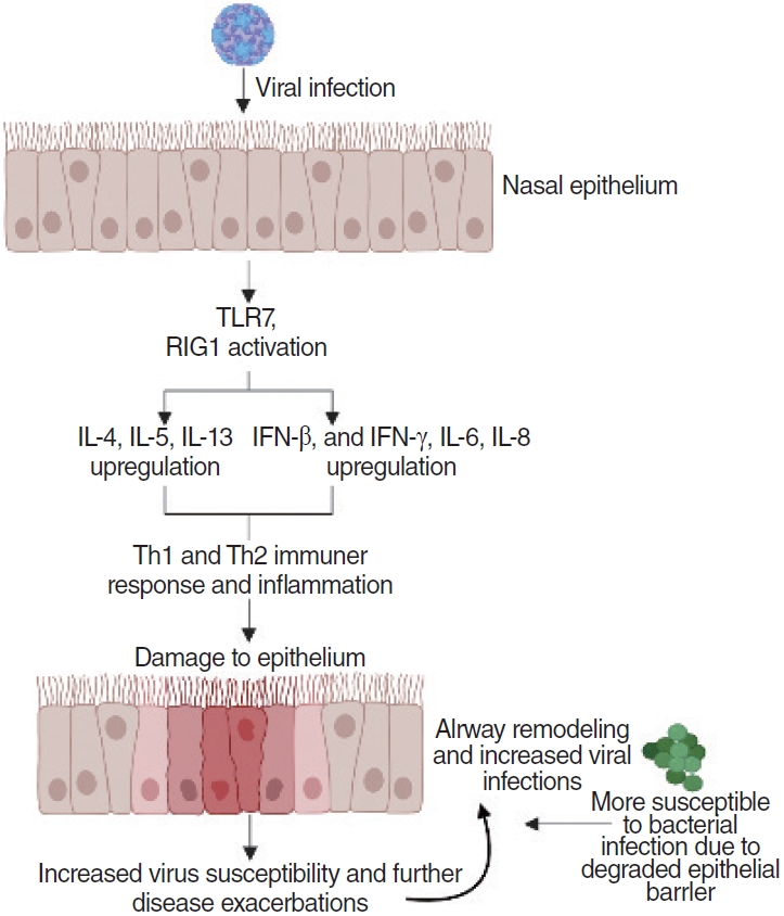
As these upper respiratory viruses infect the epithelium, they trigger immune responses that induce the release of cytokines and chemokines via intracellular sensors (TLR7 and retinoic acid-inducible gene I). Cytokines and chemokines, such as IL-6 or IFN-γ, are induced and secreted by the intracellular sensors before recruiting neutrophils and macrophages that activate the Th1 immune response [28,34,43-45]. The immune response leads to inflammation in the infected areas, which coupled with the damage from the viral infection itself and from the viral elimination by lymphocytes (e.g., Th1 cells, cytotoxic T cells), results in damage to the epithelium [60]. Continuous viral infection and inflammation cause airway remodeling of the nasal epithelium and degradation of tight junctions and adherens junctions [26,53]. Disrupted mechanical barriers and deficiencies in both the innate and acquired immune system make the sinonasal mucosa more susceptible to antigenic exposition and stimulation, leading to either side of the spectrum of chronic inflammation. This results in increased viral susceptibility of the epithelium, allowing further disease exacerbations and greater potential for bacterial infections to occur (Fig. 1). As the epithelium becomes degraded, the persistent infections and immune responses lead to CRS and CRS exacerbations [62]. Epithelial damage has been observed in CRS with nasal polyps, and genetic deficiencies or environmentally induced damage of epithelial repair mechanisms may be associated with both forms of CRS [61,63,64].

RISK FACTORS FOR VIRAL INFECTIONS IN CRS

Several risk factors can facilitate CRS infections by contributing to viral binding, entry, replication, and the immune response. Epithelial barriers are critical in preventing viral binding and entry into sinonasal epithelia. Mutations in CDHR3, the primary viral receptor for RV-C, have been associated with an increased risk for CRS and asthma [40]. One hypothesis is that the rs6963770 single-nucleotide polymorphism may result in increased RV-C binding and modulate a dysregulated immune response [65]. Age is considered a risk factor for RSV infection, as high rates of serious RSV infections and hospitalizations are observed among infants. Additionally, the presence of underlying conditions such as prematurity, congenital heart disease, immunosuppression, and cystic fibrosis all increase the risk of developing severe RSV infections [66]. Similar to RSV, PIV infections are more common and tend to be more severe in infants and young children, or elderly with compromised immune systems [67]. For influenza, a study of children in Ontario, Canada found that asthma, regardless of the severity, was a significant risk factor associated with severe disease [68].
Allergic rhinitis and asthma have a strong tendency to co-occur with CRS, suggesting a common mechanism of disease. In all three of these type 2-mediated airway disorders, the epithelial barrier is compromised. This leakiness in the epithelial barrier is hypothesized to allow enhanced viral entry through the epithelia to trigger alarmin signals including thymic stromal lymphopoietin, IL-33, and IL-13, which can trigger type 2 activation of mast cells and eosinophils. Similar mechanisms of epithelial barrier dysfunction can be seen in prolonged exposure to tobacco and air pollution, which are highly associated with CRS risk [69-72].
Aside from risk factors pertaining to an increase in CRS risk, there are risk factors for acute CRS exacerbations that increase nasal and sinus symptom severity. General health risk factors include smoking, a higher body-mass index, previous sinus surgery, and a longer CRS status, while several seasonal components such as hay fever or the winter season also increase the risk of acute CRS exacerbation [58]. Comorbid predisposing factors include asthma symptoms, impaired mucociliary clearance, and atrophic rhinitis [73-75].

CRS-RELATED VIRUSES IN CHILDREN VERSUS ADULTS

Age is a strong risk factor for CRS. Children have 3–8 viral URIs per year compared to adults who only have 2–4 URIs [76]. Male children under the age of 3 more commonly contract respiratory illnesses than female children of a similar age, while the opposite is true as their ages progress [77,78]. Comorbid conditions such as allergic rhinitis were found in 36%–60% of pediatric patients with CRS [79-81]. Khoo et al. [82] found that asthma and wheezing exacerbations in children were more prevalent at younger ages. RV-C was the most frequently identified viral pathogen in these children, and several viruses including RSV, PIV, and influenza virus were also detected.
Certain viruses are more predominant in certain age groups. Interestingly, the studies examined in this review revealed that RSV and PIV are notably more prevalent in certain age groups. For example, children and the elderly are well documented as being more susceptible to RSV infection than adolescents or adults. Immature immune systems or low lymphocyte counts in infants and young children, as well as low levels of RSV-neutralizing antibodies in patients over 65, are factors that cause these age groups to be more susceptible to infection [83,84]. Furthermore, RSV pathogenesis differs in children and adults. For example, most adults and elderly people infected with RSV show symptoms similar to influenza infection, while infants and young children with RSV infections often progress to lower respiratory tract infections and wheezing [85-87]. Additionally, PIV infection often causes URIs in most healthy young adults, but more frequently leads to severe symptoms and lower respiratory illnesses in young children. Similarly to RSV, PIV infection is one of the leading causes of acute respiratory tract infections in young children under the age of 5, accounting for approximately 17% of hospitalizations [67,88,89].

CONCLUSION

In summary, CRS affects millions of people worldwide and poses a significant financial burden. Therefore, understanding the mechanisms of infection that drive its pathology is important. In order to devise effective therapies for patients with CRS, understanding the viruses, their mechanisms of infections, and their immune responses is crucial. RVs are frequently isolated in patients with CRS. RSV, PIV, and influenza virus are also isolated in patients with CRS. These four viruses have many similarities such as targeting epithelial airway cells and being RNA viruses. However, the prevalence, receptor type, and immune response vary from virus to virus.
RVs are the most widely and thoroughly studied viruses in terms of CRS specifically, and even in terms of URI and acute sinusitis. In the future, we hope to see more studies that detail the immune response of upper respiratory tract infections and CRS due to RSV, PIV, and influenza virus. Additionally, we hope to see more longitudinal studies that follow infants and young children infected with serious respiratory viruses and how those infections can contribute to the onset of more serious cases of URI, such as CRS, later in adulthood. Throughout this review, we also noticed a scarcity of papers on pediatric CRS. Research indicates that this time point is critical in understanding the development and onset of adult CRS. Thus, further studies are necessary to be better able to target and create new therapies.

HIGHLIGHTS

▪ The four most commonly isolated viruses in patients with chronic rhinosinusitis are rhinovirus, parainfluenza virus, influenza virus, and respiratory syncytial virus.
▪ Viral infection in the upper airways has been shown to degrade epithelial barrier function, and rhinovirus infection has been specifically shown to degrade tight junction and adherens junction components.
▪ Age is strongly associated with chronic rhinosinusitis risk.
▪ Viral infections linked with chronic rhinosinusitis are more prevalent in infants and children than in adults.

CONFLICT OF INTEREST

No potential conflict of interest relevant to this article was reported.

Notes

Author contributions

Conceptualization: all authors. Data curation: HSL, SJV. Formal Analysis: all authors. Methodology: all authors. Project Administration: EHC. Writing–original draft: HSL, SJV. Writing–review & editing: EHC.

Fig. 1.
As a virus infects the upper airway epithelial cells, it activates Toll-like receptor 7 (TLR7) and retinoic acid-inducible gene I (RIG-1). These receptors induce the release of type I and type III interferons (IFNs), as well as interleukin (IL)-6 and IL-8 and other cytokines, to promote a Th1 immune response. A Th2 immune response is also induced through the production of IL-4, IL-5, IL-13, and other cytokines. The immune response creates inflammation and airway remodeling. Prolonged inflammation results in airway remodeling, which contributes to chronic rhinosinusitis (CRS) due to disrupted epithelial barrier function. As the epithelium is weak and damaged, viral susceptibility increases, resulting in further CRS and upper respiratory disease exacerbations. Additionally, the environment that results from this immune response also creates a suitable environment for bacterial infection, as the epithelial barrier is weak [28,34,43-45,61,62]. The figure was created with BioRender.com.
ceo-2022-01004f1.jpg
Table 1.
Characteristics of URI progression
Type of disease URIa) ARSb) CRSc)
Length 7–11 day (up to 14) >10 day >12 wk
Symptom Stuffy and runny nose, mild cough, watery eyes, sneezing, low-grade fever, yellow/ green nasal discharge, headache, mild fatigue Thick yellow/green mucus in the nose, facial pain and tenderness especially in the eyes, cheeks, or nose, PND, nose congestion Increased facial pain, PND, reduced sense of smell or taste, nose congestion, nasal inflammation
Endoscopy Yellow/green mucus Thick mucus Swelling and polyps
Radiology Mucosal thickening <4 mm or absence of mucosal thickening Mucosal thickening >4 mm, obstruction of osteomeatal complexes Polyps and sinus obstruction, mucosal thickening
Severity Mild Moderate Severe
Treatment Supportive (e.g., saline nasal sprays) Pain relievers, nasal steroids, decongestants, antibiotics Oral steroids, functional endoscopic sinus surgery, biologic therapies

URI, upper respiratory infection; ARS, acute rhinosinusitis; CRS, chronic sinusitis; PND, postnasal drainage.

a) The mildest form of sinus infection, lasts for 7–11 days, while ARS and CRS are more severe.

b) ARS is considered moderate, and its diagnosis requires the persistence of symptoms beyond 10 days, with a failure of improvement for at least 10 days.

c) CRS is described as severe, and is characterized by the presence of symptoms for at least 12 weeks [1,5-7].

Table 2.
Characteristics of viruses associated with CRS
Virus Characteristics Receptor Location of receptor Immune response & pathway
Rhinovirus Most common virus isolated in patients with CRS [4]. ICAM-1 and LDLR for RV-A and RV-B [18,19] ICAM-1 is expressed in most tis- sues at low levels, particularly in endothelial cells [24]. Induced expression of CXCL9, CXCL11, IP-10, and RANTES [25];
Three distinct subtypes: RV-A, RV-B, and RV-C, with RV-C being the most severe [10] CDHR3 for RV-C [21] CDHR3 is specifically expressed in ciliated epithelial airway cells [21]. Degrades tight junction and adherens junction components [26]
Influenza Causes destruction of airway epithelial cells [10] α2,6-Type receptors [27] Airway epithelial cells [27] Increased levels of IL-6, IL-8, TNF-α, IL-10, and IFN-γ [28]
RSV Common cause of respiratory infection in children [29] NCL, IGF1R, CX3CR1 [30,31] IGF1R can be expressed in lung epithelium [30]. Age-dependent immune response: IL-33 increased in neonatal mice, but not in adult mice [32,33].
CX3CR1 is expressed in immune cells and in epithelial cells [31].
PIV Primarily affects young children [34] Interaction between HN and SA-containing receptor on cell surfaces: α2-3-linked SAs, and α2-8-linked SAs [35,36] Cell surface, airway epithelial cells [35] IL-1β, IL-6, TNF-α, IL-1ra, IFN-γ, IL-2, IL-4, IL-5, IL-10, G-CSF, GM-CSF, IL-8, IP-10, eotaxin, RANTES, PDGF-BB, and VEGF [34]

Despite having different receptors, these four viruses frequently isolated in CRS patients are all expressed in epithelial cells.

CRS, chronic rhinosinusitis; RV, rhinovirus; ICAM-1, intercellular adhesion molecule 1; LDLR, low-density lipoprotein receptor; RANTES, regulated upon activation, normal T cell expressed and presumably secreted; IL, interleukin; TNF, tumor necrosis factor; IFN, interferon; RSV, respiratory syncytial virus; NCL, nucleolin; IGF1R, insulin-like growth factor 1 receptor; CX3CR, C-X3-C motif chemokine receptor; PIV, parainfluenza virus; HN, hemagglutinin-neuraminidase; SA, sialic acid; G-CSF, granulocyte colony-stimulating factor; GM-CSF, granulocyte-macrophage colony-stimulating factor; IP-10, interferon gamma-induced protein 10; PDGF, platelet-derived growth factor; VEGF, vascular endothelial growth factor.

REFERENCES

1. Rosenfeld RM, Piccirillo JF, Chandrasekhar SS, Brook I, Ashok Kumar K, Kramper M, et al. Clinical practice guideline (update): adult sinusitis. Otolaryngol Head Neck Surg. 2015 Apr;152(2 Suppl):S1-39.
crossref pdf
2. Caulley L, Thavorn K, Rudmik L, Cameron C, Kilty SJ. Direct costs of adult chronic rhinosinusitis by using 4 methods of estimation: results of the US Medical Expenditure Panel Survey. J Allergy Clin Immunol. 2015 Dec;136(6):1517-22.
crossref pmid
3. Halawi AM, Smith SS, Chandra RK. Chronic rhinosinusitis: epidemiology and cost. Allergy Asthma Proc. 2013 Jul-Aug;34(4):328-34.
crossref pmid
4. Cho GS, Moon BJ, Lee BJ, Gong CH, Kim NH, Kim YS, et al. High rates of detection of respiratory viruses in the nasal washes and mucosae of patients with chronic rhinosinusitis. J Clin Microbiol. 2013 Mar;51(3):979-84.
crossref pmid pmc pdf
5. Sonkens JW, Harnsberger HR, Blanch GM, Babbel RW, Hunt S. The impact of screening sinus CT on the planning of functional endoscopic sinus surgery. Otolaryngol Head Neck Surg. 1991 Dec;105(6):802-13.
crossref pmid pdf
6. Kamalian S, Avery L, Lev MH, Schaefer PW, Curtin HD, Kamalian S. Nontraumatic head and neck emergencies. Radiographics. 2019 Oct;39(6):1808-23.
crossref pmid
7. Shaikh N, Hoberman A, Kearney DH, Colborn DK, Kurs-Lasky M, Jeong JH, et al. Signs and symptoms that differentiate acute sinusitis from viral upper respiratory tract infection. Pediatr Infect Dis J. 2013 Oct;32(10):1061-5.
crossref pmid pmc
8. Ramadan HH, Farr RW, Wetmore SJ. Adenovirus and respiratory syncytial virus in chronic sinusitis using polymerase chain reaction. Laryngoscope. 1997 Jul;107(7):923-5.
crossref pmid
9. Abshirini H, Makvandi M, Seyyed Ashrafi M, Hamidifard M, Saki N. Prevalence of rhinovirus and respiratory syncytial virus among patients with chronic rhinosinusitis. Jundishapur J Microbiol. 2015 Mar;8(3):e20068.
crossref pmid pmc
10. Jacobs SE, Lamson DM, St George K, Walsh TJ. Human rhinoviruses. Clin Microbiol Rev. 2013 Jan;26(1):135-62.
crossref pmid pmc pdf
11. Willis AL, Calton JB, Calton J, Kim AS, Lee R, Torabzadeh E, et al. RV-C infections result in greater clinical symptoms and epithelial responses compared to RV-A infections in patients with CRS. Allergy. 2020 Dec;75(12):3264-7.
crossref pmid pdf
12. Ryu G, Shin HW. SARS-CoV-2 infection of airway epithelial cells. Immune Netw. 2021 Mar;21(1):e3.
crossref pmid pmc pdf
13. Chang EH, Willis AL, Romanoski CE, Cusanovich DA, Pouladi N, Li J, et al. Rhinovirus infections in individuals with asthma increase ACE2 expression and cytokine pathways implicated in COVID-19. Am J Respir Crit Care Med. 2020 Sep;202(5):753-5.
crossref pmid pmc
14. Onabajo OO, Banday AR, Stanifer ML, Yan W, Obajemu A, Santer DM, et al. Interferons and viruses induce a novel truncated ACE2 isoform and not the full-length SARS-CoV-2 receptor. Nat Genet. 2020 Dec;52(12):1283-93.
crossref pmid pmc pdf
15. Parma V, Ohla K, Veldhuizen MG, Niv MY, Kelly CE, Bakke AJ, et al. More than smell-COVID-19 is associated with severe impairment of smell, taste, and chemesthesis. Chem Senses. 2020 Oct;45(7):609-22.
pmid
16. Wang H, Song J, Pan L, Yao Y, Deng YK, Wang ZC, et al. The characterization of chronic rhinosinusitis in hospitalized patients with COVID-19. J Allergy Clin Immunol Pract. 2020 Nov-Dec;8(10):3597-9.
crossref pmid pmc
17. Poltronieri P, Sun B, Mallardo M. RNA viruses: RNA roles in pathogenesis, coreplication and viral load. Curr Genomics. 2015 Oct;16(5):327-35.
crossref pmid pmc
18. Bochkov YA, Gern JE. Rhinoviruses and their receptors: implications for allergic disease. Curr Allergy Asthma Rep. 2016 Apr;16(4):30.
crossref pmid pmc pdf
19. Abraham G, Colonno RJ. Many rhinovirus serotypes share the same cellular receptor. J Virol. 1984 Aug;51(2):340-5.
crossref pmid pmc pdf
20. Palmenberg AC, Gern JE. Classification and evolution of human rhinoviruses. Methods Mol Biol. 2015;1221:1-10.
crossref pmid pmc
21. Bochkov YA, Watters K, Ashraf S, Griggs TF, Devries MK, Jackson DJ, et al. Cadherin-related family member 3, a childhood asthma susceptibility gene product, mediates rhinovirus C binding and replication. Proc Natl Acad Sci U S A. 2015 Apr;112(17):5485-90.
crossref pmid pmc
22. Wiesolek HL, Bui TM, Lee JJ, Dalal P, Finkielsztein A, Batra A, et al. Intercellular adhesion molecule 1 functions as an efferocytosis receptor in inflammatory macrophages. Am J Pathol. 2020 Apr;190(4):874-85.
crossref pmid pmc
23. Stewart MP, Cabanas C, Hogg N. T cell adhesion to intercellular adhesion molecule-1 (ICAM-1) is controlled by cell spreading and the activation of integrin LFA-1. J Immunol. 1996 Mar;156(5):1810-7.
crossref pmid pdf
24. Martz E. Lymphocyte function-associated antigen 1 (LFA-1). In: Delves PJ, editor. Encyclopedia of immunology. 2nd ed. San Diego (CA): Academic Press; 1998. p. 1607-12.

25. Tan KS, Ong HH, Yan Y, Liu J, Li C, Ong YK, et al. In vitro model of fully differentiated human nasal epithelial cells infected with rhinovirus reveals epithelium-initiated immune responses. J Infect Dis. 2018 Mar;217(6):906-15.
crossref pmid
26. Yeo NK, Jang YJ. Rhinovirus infection-induced alteration of tight junction and adherens junction components in human nasal epithelial cells. Laryngoscope. 2010 Feb;120(2):346-52.
crossref pmid pdf
27. Garcia-Sastre A. Influenza virus receptor specificity: disease and transmission. Am J Pathol. 2010 Apr;176(4):1584-5.
pmid pmc
28. Hayden FG, Fritz R, Lobo MC, Alvord W, Strober W, Straus SE. Local and systemic cytokine responses during experimental human influenza A virus infection: relation to symptom formation and host defense. J Clin Invest. 1998 Feb;101(3):643-9.
crossref pmid pmc
29. Bermejo-Martin JF, Garcia-Arevalo MC, De Lejarazu RO, Ardura J, Eiros JM, Alonso A, et al. Predominance of Th2 cytokines, CXC chemokines and innate immunity mediators at the mucosal level during severe respiratory syncytial virus infection in children. Eur Cytokine Netw. 2007 Sep;18(3):162-7.
pmid
30. Griffiths CD, Bilawchuk LM, McDonough JE, Jamieson KC, Elawar F, Cen Y, et al. IGF1R is an entry receptor for respiratory syncytial virus. Nature. 2020 Jul;583(7817):615-9.
crossref pmid pdf
31. Anderson CS, Chu CY, Wang Q, Mereness JA, Ren Y, Donlon K, et al. CX3CR1 as a respiratory syncytial virus receptor in pediatric human lung. Pediatr Res. 2020 Apr;87(5):862-7.
crossref pmid pdf
32. Hijano DR, Vu LD, Kauvar LM, Tripp RA, Polack FP, Cormier SA. Role of type i interferon (IFN) in the respiratory syncytial virus (RSV) immune response and disease severity. Front Immunol. 2019 Mar;10:566.
crossref pmid pmc
33. Saravia J, You D, Shrestha B, Jaligama S, Siefker D, Lee GI, et al. Respiratory syncytial virus disease is mediated by age-variable IL-33. PLoS Pathog. 2015 Oct;11(10):e1005217.
crossref pmid pmc
34. Yoshizumi M, Kimura H, Okayama Y, Nishina A, Noda M, Tsukagoshi H, et al. Relationships between cytokine profiles and signaling pathways (IκB Kinase and p38 MAPK) in parainfluenza virus-infected lung fibroblasts. Front Microbiol. 2010 Nov;1:124.
crossref pmid pmc
35. Ah-Tye C, Schwartz S, Huberman K, Carlin E, Moscona A. Virus-receptor interactions of human parainfluenza viruses types 1, 2 and 3. Microb Pathog. 1999 Nov;27(5):329-36.
crossref pmid
36. Alymova IV, Portner A, Mishin VP, McCullers JA, Freiden P, Taylor GL. Receptor-binding specificity of the human parainfluenza virus type 1 hemagglutinin-neuraminidase glycoprotein. Glycobiology. 2012 Feb;22(2):174-80.
crossref pmid
37. Vignola AM, Chanez P, Campbell AM, Pinel AM, Bousquet J, Michel FB, et al. Quantification and localization of HLA-DR and intercellular adhesion molecule-1 (ICAM-1) molecules on bronchial epithelial cells of asthmatics using confocal microscopy. Clin Exp Immunol. 1994 Apr;96(1):104-9.
crossref pmid pmc pdf
38. Hubbard AK, Rothlein R. Intercellular adhesion molecule-1 (ICAM-1) expression and cell signaling cascades. Free Radic Biol Med. 2000 May;28(9):1379-86.
crossref pmid
39. Go GW, Mani A. Low-density lipoprotein receptor (LDLR) family orchestrates cholesterol homeostasis. Yale J Biol Med. 2012 Mar;85(1):19-28.
pmid pmc
40. Bonnelykke K, Sleiman P, Nielsen K, Kreiner-Moller E, Mercader JM, Belgrave D, et al. A genome-wide association study identifies CDHR3 as a susceptibility locus for early childhood asthma with severe exacerbations. Nat Genet. 2014 Jan;46(1):51-5.
crossref pmid pdf
41. Berger A. Th1 and Th2 responses: what are they. BMJ. 2000 Aug;321(7258):424.
pmid
42. Konnecke M, Burmeister M, Pries R, Boscke R, Bruchhage KL, Ungefroren H, et al. Epithelial-mesenchymal transition in chronic rhinosinusitis: differences revealed between epithelial cells from nasal polyps and inferior turbinates. Arch Immunol Ther Exp (Warsz). 2017 Apr;65(2):157-73.
crossref pmid pdf
43. Lavoie TB, Kalie E, Crisafulli-Cabatu S, Abramovich R, DiGioia G, Moolchan K, et al. Binding and activity of all human alpha interferon subtypes. Cytokine. 2011 Nov;56(2):282-9.
crossref pmid
44. Jaks E, Gavutis M, Uze G, Martal J, Piehler J. Differential receptor subunit affinities of type I interferons govern differential signal activation. J Mol Biol. 2007 Feb;366(2):525-39.
crossref pmid
45. Donnelly RP, Sheikh F, Kotenko SV, Dickensheets H. The expanded family of class II cytokines that share the IL-10 receptor-2 (IL-10R2) chain. J Leukoc Biol. 2004 Aug;76(2):314-21.
crossref pmid pdf
46. Briscoe J, Guschin D, Rogers NC, Watling D, Muller M, Horn F, et al. JAKs, STATs and signal transduction in response to the interferons and other cytokines. Philos Trans R Soc Lond B Biol Sci. 1996 Feb;351(1336):167-71.
crossref pmid pdf
47. Kerr IM, Costa-Pereira AP, Lillemeier BF, Strobl B. Of JAKs, STATs, blind watchmakers, jeeps and trains. FEBS Lett. 2003 Jul;546(1):1-5.
crossref pmid pdf
48. Karpusas M, Nolte M, Benton CB, Meier W, Lipscomb WN, Goelz S. The crystal structure of human interferon beta at 2.2-A resolution. Proc Natl Acad Sci U S A. 1997 Oct;94(22):11813-8.
pmid pmc
49. Klaus W, Gsell B, Labhardt AM, Wipf B, Senn H. The three-dimensional high resolution structure of human interferon alpha-2a determined by heteronuclear NMR spectroscopy in solution. J Mol Biol. 1997 Dec;274(4):661-75.
pmid
50. Gad HH, Dellgren C, Hamming OJ, Vends S, Paludan SR, Hartmann R. Interferon-lambda is functionally an interferon but structurally related to the interleukin-10 family. J Biol Chem. 2009 Jul;284(31):20869-75.
pmid pmc
51. Kim JH, Kim YS, Cho GS, Kim NH, Gong CH, Lee BJ, et al. Human rhinovirus-induced proinflammatory cytokine and interferon-β responses in nasal epithelial cells from chronic rhinosinusitis patients. Allergy Asthma Immunol Res. 2015 Sep;7(5):489-96.
crossref pmid pmc pdf
52. Nakagome K, Bochkov YA, Ashraf S, Brockman-Schneider RA, Evans MD, Pasic TR, et al. Effects of rhinovirus species on viral replication and cytokine production. J Allergy Clin Immunol. 2014 Aug;134(2):332-41.
crossref pmid pmc
53. Hulse KE. Immune mechanisms of chronic rhinosinusitis. Curr Allergy Asthma Rep. 2016 Jan;16(1):1.
crossref pmid pmc pdf
54. Pinto RA, Arredondo SM, Bono MR, Gaggero AA, Diaz PV. T helper 1/T helper 2 cytokine imbalance in respiratory syncytial virus infection is associated with increased endogenous plasma cortisol. Pediatrics. 2006 May;117(5):e878-86.
crossref pmid pdf
55. Hattori S, Shimojo N, Mashimo T, Inoue Y, Ono Y, Kohno Y, et al. Relationship between RANTES polymorphisms and respiratory syncytial virus bronchiolitis in a Japanese infant population. Jpn J Infect Dis. 2011;64(3):242-5.
crossref pmid
56. Semple MG, Dankert HM, Ebrahimi B, Correia JB, Booth JA, Stewart JP, et al. Severe respiratory syncytial virus bronchiolitis in infants is associated with reduced airway interferon gamma and substance P. PLoS One. 2007 Oct;2(10):e1038.
crossref pmid pmc
57. Ye Q, Shao WX, Shang SQ, Pan YX, Shen HQ, Chen XJ. Epidemiological characteristics and immune status of children with respiratory syncytial virus. J Med Virol. 2015 Feb;87(2):323-9.
crossref pmid
58. Kristjansson S, Bjarnarson SP, Wennergren G, Palsdottir AH, Arnadottir T, Haraldsson A, et al. Respiratory syncytial virus and other respiratory viruses during the first 3 months of life promote a local TH2-like response. J Allergy Clin Immunol. 2005 Oct;116(4):805-11.
crossref pmid
59. Skoner DP, Gentile DA, Patel A, Doyle WJ. Evidence for cytokine mediation of disease expression in adults experimentally infected with influenza A virus. J Infect Dis. 1999 Jul;180(1):10-4.
crossref pmid
60. Eloy P, Poirrier AL, De Dorlodot C, Van Zele T, Watelet JB, Bertrand B. Actual concepts in rhinosinusitis: a review of clinical presentations, inflammatory pathways, cytokine profiles, remodeling, and management. Curr Allergy Asthma Rep. 2011 Apr;11(2):146-62.
crossref pmid pmc pdf
61. Ramshaw IA, Ramsay AJ, Karupiah G, Rolph MS, Mahalingam S, Ruby JC. Cytokines and immunity to viral infections. Immunol Rev. 1997 Oct;159:119-35.
crossref pmid
62. Yan Y, Gordon WM, Wang DY. Nasal epithelial repair and remodeling in physical injury, infection, and inflammatory diseases. Curr Opin Otolaryngol Head Neck Surg. 2013 Jun;21(3):263-70.
crossref pmid
63. Tan KS, Yan Y, Ong HH, Chow VT, Shi L, Wang DY. Impact of respiratory virus infections in exacerbation of acute and chronic rhinosinusitis. Curr Allergy Asthma Rep. 2017 Apr;17(4):24.
crossref pmid pmc pdf
64. Hoggard M, Wagner Mackenzie B, Jain R, Taylor MW, Biswas K, Douglas RG. Chronic rhinosinusitis and the evolving understanding of microbial ecology in chronic inflammatory mucosal disease. Clin Microbiol Rev. 2017 Jan;30(1):321-48.
crossref pmid pdf
65. Basnet S, Bochkov YA, Brockman-Schneider RA, Kuipers I, Aesif SW, Jackson DJ, et al. CDHR3 asthma-risk genotype affects susceptibility of airway epithelium to rhinovirus C infections. Am J Respir Cell Mol Biol. 2019 Oct;61(4):450-8.
crossref pmid pmc
66. Aujard Y, Fauroux B. Risk factors for severe respiratory syncytial virus infection in infants. Respir Med. 2002 Apr;96 Suppl B:S9-14.
crossref pmid
67. Reed G, Jewett PH, Thompson J, Tollefson S, Wright PF. Epidemiology and clinical impact of parainfluenza virus infections in otherwise healthy infants and young children <5 years old. J Infect Dis. 1997 Apr;175(4):807-13.
crossref pmid
68. Meier CR, Napalkov PN, Wegmuller Y, Jefferson T, Jick H. Population-based study on incidence, risk factors, clinical complications and drug utilisation associated with influenza in the United Kingdom. Eur J Clin Microbiol Infect Dis. 2000 Nov;19(11):834-42.
crossref pmid pdf
69. Hastan D, Fokkens WJ, Bachert C, Newson RB, Bislimovska J, Bockelbrink A, et al. Chronic rhinosinusitis in Europe: an underestimated disease: a GA²LEN study. Allergy. 2011 Sep;66(9):1216-23.
pmid
70. Wolf C. Urban air pollution and health: an ecological study of chronic rhinosinusitis in Cologne, Germany. Health Place. 2002 Jun;8(2):129-39.
crossref pmid
71. Bhattacharyya N. Air quality influences the prevalence of hay fever and sinusitis. Laryngoscope. 2009 Mar;119(3):429-33.
crossref pmid
72. Thilsing T, Rasmussen J, Lange B, Kjeldsen AD, Al-Kalemji A, Baelum J. Chronic rhinosinusitis and occupational risk factors among 20- to 75-year-old Danes: a GA(2) LEN-based study. Am J Ind Med. 2012 Nov;55(11):1037-43.
crossref pmid
73. Kuiper JR, Hirsch AG, Bandeen-Roche K, Sundaresan AS, Tan BK, Schleimer RP, et al. Prevalence, severity, and risk factors for acute exacerbations of nasal and sinus symptoms by chronic rhinosinusitis status. Allergy. 2018 Jun;73(6):1244-53.
crossref pmid pdf
74. Hafner B, Davris S, Riechelmann H, Mann WJ, Amedee RG. Endonasal sinus surgery improves mucociliary transport in severe chronic sinusitis. Am J Rhinol. 1997 Jul-Aug;11(4):271-4.
crossref pmid pdf
75. Dutta M, Ghatak S. Acute exacerbation of chronic rhinosinusitis (AECRS) with orbital complications in an atrophic rhinitis patient: a mere co-incidence. J Clin Diagn Res. 2013 Dec;7(12):2973-5.
crossref pmid pmc
76. Hamilos DL. Pediatric chronic rhinosinusitis. Am J Rhinol Allergy. 2015 Nov-Dec;29(6):414-20.
crossref pmid pdf
77. Wald ER. Sinusitis in children. N Engl J Med. 1992 Jan;326(5):319-23.
crossref pmid
78. Monto AS, Ullman BM. Acute respiratory illness in an American community: the Tecumseh study. JAMA. 1974 Jan;227(2):164-9.
crossref pmid
79. Shapiro GG, Virant FS, Furukawa CT, Pierson WE, Bierman CW. Immunologic defects in patients with refractory sinusitis. Pediatrics. 1991 Mar;87(3):311-6.
crossref pmid pdf
80. Kogutt MS, Swischuk LE. Diagnosis of sinusitis in infants and children. Pediatrics. 1973 Jul;52(1):121-4.
crossref pmid pdf
81. Rachelefsky GS, Siegel SC, Katz RM, Spector SL, Rohr AS. Chronic sinusitis in children. J Allergy Clin Immunol. 1991 Jan;87(1):219.

82. Khoo SK, Read J, Franks K, Zhang G, Bizzintino J, Coleman L, et al. Upper airway cell transcriptomics identify a major new immunological phenotype with strong clinical correlates in young children with acute wheezing. J Immunol. 2019 Mar;202(6):1845-58.
crossref pmid
83. Hall CB, Powell KR, MacDonald NE, Gala CL, Menegus ME, Suffin SC, et al. Respiratory syncytial viral infection in children with compromised immune function. N Engl J Med. 1986 Jul;315(2):77-81.
crossref pmid
84. Berbers G, Mollema L, van der Klis F, den Hartog G, Schepp R. Antibody responses to respiratory syncytial virus: a cross-sectional serosurveillance study in the Dutch population focusing on infants younger than 2 years. J Infect Dis. 2021 Jul;224(2):269-78.
crossref pmid pdf
85. Lozano R, Naghavi M, Foreman K, Lim S, Shibuya K, Aboyans V, et al. Global and regional mortality from 235 causes of death for 20 age groups in 1990 and 2010: a systematic analysis for the Global Burden of Disease Study 2010. Lancet. 2012 Dec;380(9859):2095-128.
pmid
86. Mochizuki H, Todokoro M, Arakawa H. RS virus-induced inflammation and the intracellular glutathione redox state in cultured human airway epithelial cells. Inflammation. 2009 Aug;32(4):252-64.
crossref pmid pdf
87. Bonville CA, Rosenberg HF, Domachowske JB. Macrophage inflammatory protein-1alpha and RANTES are present in nasal secretions during ongoing upper respiratory tract infection. Pediatr Allergy Immunol. 1999 Feb;10(1):39-44.
pmid
88. Karron RA, Collins PL. Parainfluenza viruses. In: Knipe DM, Howley PM, editors. Fields virology. 5th ed. Philadelphia (PA): Lippincott Williams & Wilkins; 2007. p. 1497-526.

89. do Carmo Debur M, Raboni SM, Flizikowski FB, Chong DC, Persicote AP, Nogueira MB, et al. Immunohistochemical assessment of respiratory viruses in necropsy samples from lethal non-pandemic seasonal respiratory infections. J Clin Pathol. 2010 Oct;63(10):930-4.
crossref pmid
Editorial Office
Korean Society of Otorhinolaryngology-Head and Neck Surgery
103-307 Park Tower officetel, Yongsan-dong 5-ga, Yongsan-gu, Seoul 04385, Korea
TEL: +82-2-711-9091   FAX: +82-2-3487-6603   E-mail: editor.eceo@gmail.com
Copyright © Korean Society of Otorhinolaryngology-Head and Neck Surgery.                 Developed in M2PI
Close layer
prev next