The Promoting Effect of Carbamide Peroxide Teeth Bleaching Gel in a Preclinical Model of Head and Neck Cancer in Hamster Buccal Pouch
Article information
Abstract
Objectives
The aim of this study was to verify the promoting effect of carbamide peroxide on dimethylbenzanthracene (DMBA)-induced carcinogenesis in the hamster buccal pouch, in order to reduce the period of latency for tumor formation.
Methods
Sixteen hamsters were randomized into two groups of eight animals each. The hamsters of the group I had their right buccal pouches treated with 0.5% DMBA and 10% carbamide peroxide teeth bleaching gel for 55 days. The animals of the group II had their right pouches treated only with DMBA. After, six animals of each group had their pouches prepared for light microscopy. Histomorphometry was performed to assess the presence of keratinization, nuclear polymorphism, pattern of invasion, number of blood vessels, and inflammatory infiltrate in the tumor front. Furthermore, the newly formed lesions were graded according the Bryne's grading system. The remaining animals had the vascular system of the pouches casted by Mercox and qualitatively analyzed by scanning electron microscopy.
Results
Histopathological analysis of the buccal pouches treated with DMBA and carbamide peroxide exhibited formation of squamous cell carcinoma well-differentiated with a high degree of malignancy in all pouches. The development of this neoplasm was associated with a significant increase in the number of blood vessels, presence of keratin pearls, and inflammatory infiltrate. The pouches of the group II showed inflammation, epithelial hyperplasia, dysplasia, and squamous cell carcinoma in only three right pouches. The analysis of the electron micrographs of the pouches chemically inducted with DBMA and carbamide peroxide reveled formation of a new vascular network characteristic of squamous cell carcinoma.
Conclusion
The protocol presented here, using DMBA associated with carbamide peroxide, shortens the period of latency to produce squamous cell carcinoma in the hamster buccal pouch, decreasing the time and costs of the experiments.
INTRODUCTION
The buccal pouch of the Syrian golden hamster (Mesocricetus auratus) is an excellent model to study progressive anatomical modifications and functional vascular alterations that occur in the mucosa during chemical carcinogenesis, because of the low cost and easy anatomical access, which can be readily everted for macroscopic follow-up and local treatment [1].
The induction model of squamous cell carcinoma with using dimethylbenzanthracene (DMBA) alone in the buccal pouch is a well-established cancer model for head and neck carcinomas and it has been used for over fifty years in carcinogenesis studies [2,3,4]. The DMBA produces tumors in this site in 16 weeks, what was firstly described by Salley [2]. Morris [5] improved and standardized this protocol for tumor induction, using DMBA at 0.5% and 5 weeks old hamsters. Recently, Heber et al. [6] have established a protocol, obtaining tumors in 90% of the buccal pouches treated with DMBA for 12 weeks.
The greatest disadvantages of this model is the intensive procedure in the laboratory, the constant handling throughout the experiment with the animal and the carcinogen [3], and the long latency period for tumor development, requiring several weeks of treatment until the formation of tumors.
It has been established that there are two stages in the DMBA-induced buccal pouch carcinogenesis: an initiation phase, induced by a carcinogen, and a phase of tumor promotion, stimulated by a promoter. These stages were first observed after using a solution of benzoyl peroxide to promote tumor in this model of squamous cell carcinoma of head and neck [7,8].
The carbamide peroxide has hydrogen peroxide as active ingredient, a substance widely used as a technique of vital teeth bleaching [9]. This peroxide is an unstable substance, which releases highly reactive free radicals that can damage the cell membranes and DNA [10,11].
It was found that hydrogen peroxide has a potentiating effect on duodenal carcinogenesis in rodents [12,13] and that increases the mitotic activity, and promotes hyperplasia in the epithelium of the hamster buccal pouch during DMBA-induced carcinogenesis. Alone, however, hydrogen peroxide failed to induce cancer in the buccal pouch and therefore it has been suggested as a promoting agent [14].
Although the increasing popularity of the teeth bleaching treatments, there are only few studies that investigate the promoting effect of carbamide and hydrogen peroxide in cancer models. In addition, the establishment of protocols using new substances as promoter agents is needed to reduce the time required to produce chemically inducted cancers, due to the long period of latency for tumor formation.
The aim of this study was to verify the promoting effect of carbamide peroxide in the DMBA-induced squamous cell carcinoma in the hamster buccal pouch, in order to reduce the period of latency for tumor formation.
MATERIALS AND METHODS
Animals and experimental design
All the experimental procedures performed in this study were carried out according with principles stated in Guide for the Care and the Use of Laboratory Animals [15] and were previously approved by the Ethics Committee on Animal Experimentation of Federal University of Pelotas (No. 23110.005503/2010-18). Sixteen male golden Syrian hamsters (M. auratus) obtained from the vivarium of Federal University of Pelotas were used in this study. At the beginning of the experiment they were four weeks old and weighing on average 70 g.
During the experiment, the animals were kept in opaque plastic cages, containing four animals each cage, on ventilated cabinets under controlled temperature (21℃±2℃) and humidity (60%-70%), respecting the light/dark cycle of 12 hours. Water and pelleted food for rodents were provided ad libitum.
The animals were randomly divided into two groups of eight animals each. The animals of the group I had their right buccal pouches treated with DMBA and carbamide peroxide for 55 days. The group II was treated only with DMBA for the same period of time. The left buccal pouches of both groups were not submitted to tumor induction, being considered as a control.
Tumors were induced by application of DMBA (Sigma Chemical, St. Louis, MO, USA) diluted to 0.5% in acetone, in association with carbamide peroxide teeth bleaching gel at 10% (Opalescence 10%; Ultradent Products Inc., South Jordan, UT, USA) in the animals of the group I. The carcinogen and promoter gel were applied by painting the fundus of the right pouch with a number 4 paintbrush. In the group I the applications were performed five times per week for a period totaling 55 days. The applications occurred on alternate days, three days per week for DMBA and two days per week for the promoting agent. The animals of the group II had the carbamide peroxide application skipped, having their right buccal pouches exclusively treated with DMBA three times per week. In order to observe tumor development and growth, right buccal pouches were evaluated weekly by visual inspection before the application of the chemical agents.
Histopathological and morphological analysis
For histopathological analysis, six animals from each group were euthanized by intraperitoneal injection of phenobarbital sodium (0.15 mg/g, Unifenobarb, União Química, São Paulo, Brazil) after tumor induction. The pouches were dissected and fixed in formalin solution 10%. They were then immersed in Karnovsky for 24 hours, dehydrated in ethanol and embedded in paraffin. 4 µm thick sections were obtained using a 2045 Multicut rotative microtome (Reichert-Jung, Nußloch, Germany), stained with hematoxylin and eosin (H&E), and mounted on microscope slides with Entellan (Merck, Darmstadt, Germany).
Using a ×20 objective lens, 100 images were taken for each group using the light microscope Eclipse E200 (Nikon, Tokyo, Japan) connected to the digital camera Moticam 5 (Motic Inc., Xiamen, China). A grid containing 200 points was superimposed over the images to quantify the presence of keratinization, nuclear polymorphism, pattern of invasion, number of blood vessels, and inflammatory infiltrate in areas considered invasive front of the tumor, located in the most aggressive areas of the carcinoma.
The images were analyzed by the software Image Pro-plus 4.5 (Media Cybernetics, Silver Spring, MD, USA), and 2,000 points were counted per animal. In this analysis it was calculated the amount of points that occurred over vessels, epithelial cell layer, layer of keratin, keratin pearls and inflammatory infiltrate.
For morphological analysis, the malignancy grading system proposed by Bryne [16] was followed. This system is based on four morphological parameters in the region of the invasive front of the tumor, represented by (1) degree of keratinization, (2) nuclear polymorphism, (3) pattern of invasion, and (4) inflammatory cell infiltration. All samples were examined in a magnification of ×20 and ×40, and each of the analyzed parameters received a score ranging from 1 to 4. For each of the four parameters analyzed, a sum was made and then performed a division by 4, thereby obtaining an average in each case evaluated. After obtaining this average, each case was classified as low or high-grade malignancy, following criteria described by Anneroth et al. [17], in which the score between 1.0 and 2.5 indicates a low degree of malignancy and between 2.6 and 4.0 indicates a high degree of malignancy. All morphological and histomorphometric analysis were performed by a blinded examiner.
Data were expressed as measures of central mean±SD. The normal distribution of data was verified using the Kolmogorov-Smirnov test. Based on this test, the data were analyzed using analysis of variance (ANOVA) one-way unifactorial variance, followed by Student-Newman-Keuls' test for multiple comparisons. In all analysis significance level of 5% was used. All tests were performed in GraphPad Prism 5.0 (GraphPad Software Inc., San Diego, CA, USA).
Analysis of the tumor vasculature
Two animals of each group were anesthetized by intraperitoneal injection of ketamine hydrochloride (150 mg/kg) (Dopamin, Laboratório Agribrands, São Paulo, Brazil) and xylazine (10 mg/kg; Anasedan, Laboratório Agribrands). After the anesthesia, the vascular networks of these animals were submitted to the corrosion casting technique adapted from the method previously described by Lametschwandtner et al. [18] using Mercox resin (Ladd Research Industries, Burlington, VT, USA).
The vascular casts were fixed in aluminum stubs with double-faced carbon tape and sputter coated by carbon (SCD 005 Sputter Coatter; Bal-Tec, Balzers, Germany) and gold (CEA 035 Sputter Coatter; Bal-Tec). The specimens were analyzed by the scanning electron microscopy (SEM) Phillips XL 30 (Phillips, Eindhoven, The Netherlands).
The analysis of electron micrographs was performed descriptively according to Konerding [19], evaluating the following aspects: pattern of organization of the vascular network, course of the vessels, identification of the blood vessel, frequency of vascular branches and figures of angiogenesis.
RESULTS
All the right buccal pouches treated for 55 days with DMBA associated with carbamide peroxide (group I) presented exophydic tumors, which were observed by visual examination of these pouches. In the control pouches of both groups it was not observed macroscopic alterations throughout the experiment (Fig. 1).
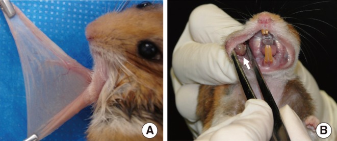
(A) Image of an everted left buccal pouch. (B) Image of a buccal pouch showing an exophytic tumor after 55 days of tumor induction with dimethylbenzanthracene and carbamide peroxide.
The histopathological analysis of the buccal pouches of the group I showed formation of squamous cell carcinoma well-differentiated in all right buccal pouches after 55 days of treatment. The animals of the group II presented in their right buccal pouches large areas with hyperplasia and inflammation. In this group, five animals presented areas of dysplasia and three of them presented squamous cell carcinoma well-differentiated. None of the changes found in the pouches submitted to the treatment were identified in the control pouches of both groups.
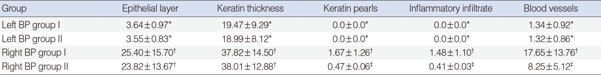
It was observed that the number of cells in the epithelial layer was significantly higher in the treated groups compared to controls pouches (P<0.05). This increase of the epithelium layers occurred primarily by invagination of epithelial cells into the underlying connective tissue. The keratin layer was significantly thicker in the treated pouches of both groups than the control pouches. These right pouches showed an increased deposition of keratin pearls in the connective tissue and epithelium (P<0.05).
The inflammatory infiltrate also presented a significant increase among the left pouches and the right pouches of the groups I and II (P<0.05). Regarding the number of blood vessels, the right buccal pouches of group I showed an increase when compared to control groups and the right pouches of the group II (P<0.05). The data described above is showed in Table 1 and the histological sections are exhibited in Fig. 2.
Influence of dimethylbenzanthracene exposure time associated with carbamide peroxide on the parameters of histoarchitecture of the hamster buccal pouch (BP)
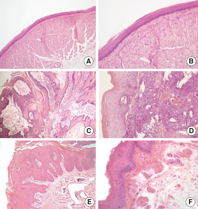
(A, B) The control pouches presented a stratified squamous orthokeratinized epithelium followed by dense connective tissue (lamina propria), striated muscle tissue and loose connective tissue. (C, D) All the right buccal pouches of the group I presented squamous cell carcinoma with a high pattern of invasion, keratin pearls and an increased vascularity. (E, F) After 55 days of exposure to dimethylbenzanthracene exclusively, there was an increase in the thickness of keratin and epithelium layers, inflammation and dysplasia (light microscopy, ×10 in the right column and ×20 in the left column, H&E stain).
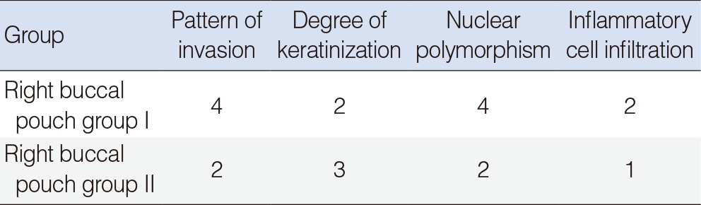
Morphological analysis of histological sections classified the groups into high and low-grade malignancy, according to the criteria established by Bryne [16]. It was observed that the buccal pouches treated only with DMBA had low-grade malignancy. In other hand, the treated pouches of group I had high-grade malignancy, mostly due to the pattern of cell invasion and increased nuclear polymorphism (Table 2). This analysis was not performed in the control groups, because they showed no alterations.
The results described above were supported by the scanning electron microscopy analysis of the vascular casts. It was observed malignant changes in the angioarchitecture of the buccal pouches.
The vascular network observed in the control buccal pouches was adequate to the physiology of the organ, presenting straight vessels, with no variation in the caliber along its course (Fig. 3A, B). In the group I, the vascular network of the treated pouches showed marked changes, associated with alterations in the vessels caliber. These blood vessels assumed glomeruloid and fingerlike aspect. Moreover, it was found an altered venous merging pattern (Fig. 3C, D), opposing the vasculature of normal tissues. The right buccal pouches of the group II showed initial changes in the course of the blood vessels, revealing the formation of a new vascular network less modified than the pouches of the group I (Fig. 3E, F).
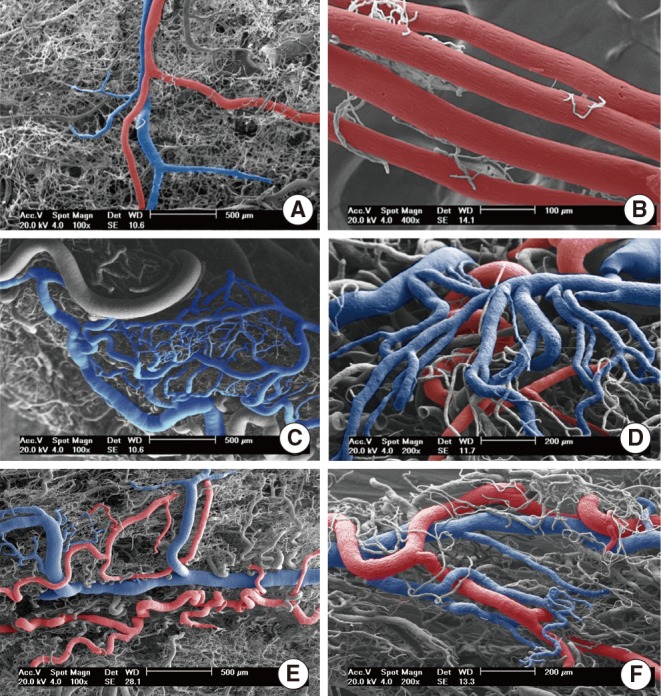
Scanning electron micrographs of vascular casts of hamster buccal pouches. (A) Left buccal pouch of group I (×100). (B) Left buccal pouch of group II (×400). (C, D) Right buccal pouch of group I (×100, ×200). (E, F) Right buccal pouch of group II (×100, ×200). Main arteries and veins are colored in red and blue consecutively.
DISCUSSION
Oral and pharyngeal cavity is the most common site of the head and neck squamous cell carcinoma. The annual estimated incidence is around 275,000 for oral and 130,300 for pharyngeal cancers and over 50% of the cases diagnosed annually are lethal. In countries like Brazil, India, Philippines, Sri Lanka, and Vietnam this type of cancer accounts for 25% of all existing cancers [20,21]. Prevention and treatment of this neoplasm requires improved animal models in which chemoprophylaxis and chemotherapy can be tested [1].
The DMBA-induced squamous cell carcinoma in hamster buccal pouch presents a close relationship between cellular and molecular events that occurred in the development of pre-malignant and malignant lesions in human beings, such as mutations and changes of expression of oncogenes and markers of cell proliferation [22]. In addition, the pouch is an excellent organ for studies of the microcirculation due to its translucent walls held by a long and continuous retractor muscle [23,24].
The establishment of a squamous cell carcinoma well-differentiated was observed in all animals after 55 days treatment with DMBA and carbamide peroxide. The application of the DMBA alone at the same conditions and period of time was insufficient to produce tumors in all animals. The development of the neoplasm in the group I was associated with a significant increase in the number of blood vessels, presence of keratin pearls, and inflammatory infiltrate when compared with the pouches treated exclusively with DMBA.
The use of the corrosion cast method associated with SEM emphasized the findings observed by light microscopy. The changes detected by light microscopy after the tumor development were correlated with morphological characteristics of the angioarchitecture presented by the pouches treated for 55 days with DMBA and carabamide peroxide.
The influence of hydrogen and carbamide peroxide on the model of DMBA-induced buccal pouch carcinoma was tested consecutively by Weitzman et al. [14] and Collet et al. [25]. In these studies, the production of squamous cells carcinoma was ineffective after a period superior to 22 weeks of treatment with DMBA two times a week used alternately with hydrogen or carbamide peroxide.
The long latency period for tumor development presented by these authors, compared with the present study, may have been due to the low concentration and frequency of application of the DMBA solution used in their protocols, less than that needed for the effective production of a neoplasm as stated by Morris [5]. Heber et al. [6] observed that the reduction of frequency of DMBA application to 2 times a week produced tumors in only 47% of the animals after 8 weeks of treatment.
In the present study, the application of DMBA three times a week, as determined by Morris [5], supplemented with the application of the teeth bleaching gel twice a week, produced squamous cell carcinoma in a period of approximately 8 weeks in all pouches submitted to the treatment. The pouches treated only with DMBA presented a longer latency period, exhibiting squamous cell carcinoma solely in three of the eight right pouches where tumors were induced. Due to the result obtained here, it is possible to suggest that the carbamide peroxide could act as a promoter on DMBA-induced carcinogenesis in hamster buccal pouch.
Others substances have been used to reduce the period of latency of the DMBA-induced carcinogenesis. Lin et al. [26] and Wani et al. [27] were successful to produce tumor after DMBA initiation followed by promotion with arecaidine, one of the main constituents of betel nut. However, in both studies it was needed at least 8 weeks of DMBA induction plus 4 additional weeks for arecaidine promotion to obtain up to 100% of squamous cell carcinoma in buccal pouches. In the present work, the concomitant use of DMBA with the carbamide peroxide gel produced this neoplasm in all pouches four weeks earlier than these studies.
The reduction of the period required for tumor formation is of the great importance for chemical carcinogenesis in animal models. The recent studies involving DMBA-inducted carcinomas in this model [24,28] still use the classic protocols [2,5] for tumor induction. The method described here, associating DMBA with carbamide peroxide, could facilitate the pre-clinic studies on head and neck carcinomas, reducing the laboratory labor and the exposure of the researchers to carcinogenic agents, shortening the time necessary to perform a study, in addition to decrease the costs of experiments involving chemical carcinogenesis.
In conclusion, the use of carbamide peroxide as a promoter in the DMBA-induced squamous cell carcinoma model shortened the period of latency to develop this tumor in the hamster buccal pouch. The protocol established here can be used to produce squamous cell carcinoma well-differentiated in a period of 55 days in the hamster buccal pouch.
ACKNOWLEDGMENTS
Coordination for the Improvement of Higher Education Personnel (CAPES; funding agency of Brazilian Ministry of Education) is gratefully acknowledged for the award of a PhD scholarship to Vinícius Faccin Bampi.
Notes
No potential conflict of interest relevant to this article was reported.