A Hypopharyngeal Ductal Cyst Masquerading as a Laryngopharyngeal Reflux Disease
Article information
Abstract
The ductal cyst of the hypopharynx is a very rare tumor. We report a case of hypopharyngeal ductal cyst in a 63-year-old man presenting with globus sensation. It was removed by a laryngomicrosurgical technique, using a microdissection electrode. Masses of the hypopharynx may not always be easily visible on routine examination of the hypopharynx with flexible fiberoptic laryngoscopes. Particularly in cases of benign tumors, the diagnosis may be delayed due to a prolonged history of mild and subtle symptoms. We missed the hypopharyngeal mass at the initial presentation, but could detect the mass in the pyriform sinus with a double contrast barium swallow study. We describe the diagnostic method to detect hypopharyngeal tumors and the treatment of benign hypopharyngeal masses.
INTRODUCTION
Benign tumors of the hypopharynx are very rare, with the most common two being the fibrolipoma and leiomyoma. The ductal cysts are mucous retention cysts that develop after obstruction of the collecting ducts of the submucosal glands. Although the oral cavity or larynx are often involved, hypopharyngeal localization is extremely rare. We report a new case of a ductal cyst that developed on the lateral wall of the pyriform sinus of the hypopharynx. There was a delay in the diagnosis because no mass-like lesion was found with routine fiberoptic laryngoscopic examination.
CASE REPORT
A 64-year-old man presented with a four-year history of globus sensation. He was referred for intractable symptoms despite having taken reflux medication for two months. Despite continued medication, his symptoms developed to dysphagia, even when swallowing liquid. He denied hoarseness or dyspnea. His medical history included a transurethral resection for bladder cancer 3 years previously. He had experienced weight loss of 3 kg over the previous 2 months. On his first presentation, flexible fiberoptic laryngoscopy showed pooling saliva, but no mass-like lesion in the hypopharynx. Vocal fold mobility was intact. The patient underwent a double-contrast pharyngoesophagogram due to persistent symptoms. An ovoid submucosal mass with a smooth contour in the left pyriform sinus was revealed (Fig. 1A). Subsequently, computed tomography (CT) scans showed a 3×4 cm mass with central low attenuation in the left hypopharynx (Fig. 1B, C). With the patient under general anesthesia, the lesion was removed via direct laryngoscopy using a microdissection electrode for endoscopic electrosurgery. The mass originated from the lateral wall of the pyriform sinus. It appeared as a pedunculated mass with a smooth, well-encapsulated surface. It was removed by means of submucosal dissection with the microelectrode. The resected specimen consisted of a 2×3×4 cm cystic mass with proteinaceous fluid (Fig. 2A). Histologic examination showed a true epithelial-lined cyst consisting of cuboidal epithelium (Fig. 2B). The final diagnosis, made by a pathologist, was a ductal cyst of the hypopharynx. Postoperative recovery of the patient was uneventful. The patient was free of symptoms after one month. Five years of follow-up showed no tumor recurrence and normal swallowing.
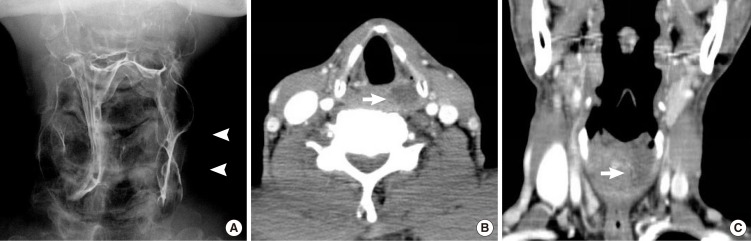
(A) Frontal view from a double-contrast pharyngoesophagogram shows a large contour defect (arrowhead) in the left pyrifrom sinus. (B, C) The axial and coronal contrast-enhanced computed tomography scans show a well-circumscribed, hypodense lesion (arrow) in the left pyriform sinus of the hypopharynx.

(A) The surgical specimen was myxoid and well-encapsulated, measuring 2×3×4 cm. (B) Microscopic examination revealed that the cyst wall was lined by cuboidal epithelial cells and had loose connective tissue in the stroma (H&E, ×200).
DISCUSSION
Benign hypopharyngeal cysts have rarely been reported in the English language literature. Therefore, the incidence of hypopharyngeal cysts is uncertain. In particular, ductal cysts of the hypopharyx are very rare. DeSanto et al. [1] reported that the ductal cyst is one of the two most common types of cysts in the larynx. Ductal cysts are caused by dilatation and obstruction of mucous gland ducts. Pathologically, the epithelium-lined cyst wall consists of a uniform layer of cuboidal, columnar, or non-keratinizing squamous epithelium. Ductal cysts usually are slow-growing and painless, appearing as a circumscribed and often fluctuant swelling. The presence or absence of symptoms in patients with benign pharyngeal masses is directly related to the size and location of these lesions.
In our case, we missed the mass at the patient's first visit. Despite intractable globus symptoms, we could not find any specific abnormality in the larynx or hypopharynx by routine fiberoptic laryngoscopy. A number of techniques have been suggested to improve the view of the hypopharynx with flexible fiberoptic laryngoscopy. One such technique is letting the patient sustain high-pitch phonation or perform a modified valsalva maneuver. Hillel and Schwartz [2] suggested performing a trumpet maneuver to fully visualize the pyriform sinus and the postcricoid region. In addition, Purser and Antippa [3] reported that manual anterosuperior traction applied to the prelaryngeal skin results in a useful view of the hypopharynx.
When nothing can be detected despite multiple laryngoscopic methods during outpatient examination in dysphagia patients, double contrast pharyngoesophagography may be useful for evaluating both the structure and the function of the hypopharynx and other organs involved in swallowing [4]. Benign lesions typically appear as small, round or ovoid, well-circumscribed, smooth-surfaced submucosal masses etched in white, best visualized on frontal views of the pharynx. These findings obviate further diagnostic workup unless symptoms are severe enough to warrant excision. CT or magnetic resonance imaging (MRI) is useful in determining the extent and location of the cyst and distinguishing it from malignant tumors. Most hypopharyngeal tumors are squamous cell carcinomas. Therefore, hypopharyngeal masses should be differentiated from malignant tumors. Our patient had a history of transitional cell carcinoma in the bladder. He underwent endoscopic excision five times for recurrent bladder cancer. Therefore, we included the metastatic mass in the differential diagnosis before surgery.
Generally, there is no reason to resect small pharyngeal retention cysts that are not causing symptoms. Larger cysts or those causing dysphagia or other symptoms need to be removed. The treatment of hypopharyngeal cysts is size-dependent [5]. The surgical removal of a small cyst is simple, and many of these cysts may be removed by endopharyngeal microsurgery using a direct laryngoscope. Surgical excision is curative. Laryngo-endoscopic microsurgery using a carbon dioxide (CO2) laser is efficient. Marsupialization may be adequate, and it may be necessary to reduce the size of a large cyst by puncturing and draining it before complete removal. We successfully removed the ductal cyst in our patient with a microdissection electrode under direct laryngoscopy. Microelectrodes are useful for resecting larygopharyngeal masses with excellent hemostasis [6]. The tissue damage is no greater than with CO2 laser injury [7].
We recommend that, in any patient with a history of intractable globus symptoms, careful physical examination with a flexible laryngoscope and radiologic studies such as double contrast pharyngoesophagogram, CT, or MRI should be performed to rule out other benign or malignant lesions.
Notes
No potential conflict of interest relevant to this article was reported.