Effect of Proparacaine in a Mouse Model of Allergic Rhinitis
Article information
Abstract
Objectives
Lidocaine, a local anaesthetic is a treatment option in uncontrolled asthma due to its immunomodulatory effects. In the present study, proparacaine (PPC), a derivative of lidocaine was examined for its therapeutic application in a mouse model of allergic rhinitis.
Methods
The mice were grouped into 4 groups: control group, allergic rhinitis (AR) group, ciclesonide (CIC) group, and PPC group. Nasal symptom scores, eosinophil counts, goblet cell counts, and mast cells counts in the nasal mucosa were measured. Serum ovalbumin (OVA)-specific immunoglobulin (Ig) E, OVA-specific IgG1, OVA-specific IgG2a, interleukin (IL)-4, IL-5, and cortisol levels were measured.
Results
Intranasal administration of PPC significantly decreased nasal symptoms, number of eosinophils, goblet cells, and mast cells in the lamina propria of the nasal mucosa. Serum OVA-specific IgE, OVA-specific IgG1, OVA-specific IgG2a was significantly higher in the AR compared with the control group. Serum level of IL-4 was significantly lower in the CIC group and PPC group in comparison with AR group. Serum IL-5 showed no significant difference among all groups. No significant difference in serum cortisol levels was observed among the 4 groups.
Conclusion
PPC appears to have a therapeutic potential in treatment of allergic rhinitis in a mouse model by reducing eosinophil, goblet cell, and mast cell infiltration in the nasal mucosa.
INTRODUCTION
Five-hundred million people worldwide suffer from allergic rhinitis (AR) and its prevalence is known to increase at an alarming rate [1]. Allergic rhinitis is a chronic inflammatory disorder caused by T cells and related T-helper (Th) 2 cytokines in which infiltration of eosinophils plays a crucial role. Glucocorticoids have been the mainstay of treatment in allergic rhinitis but long-term treatment with glucocorticoids results in many different adverse effects [2-4]. In spite of many treatments options for AR, there is a demand for novel therapeutic options which has similar anti-inflammatory effects of glucocorticoids, but with fewer side effects.
Many studies examined the underlying mechanism of lidocaine on allergic inflammation. Lidocaine is known to have significant effects on nonexcitable cells, including neutrophils, monocytes, Th2 lymphocytes, mast cells, and eosinophils [5-11]. A previous study found that local anesthetics, such as lidocaine and it analogues reduced eosinophil response to cytokines which resulted in immune modulatory effects [5]. A different study found that in a mouse model of asthma, lidocaine inhalation reduced eosinophilic inflammation, mucus production, and peribronchial fibrosis [6]. Recently, nebulized lidocaine is used on patients with asthma who were dependent on oral glucocorticoids and successfully replaced oral glucocorticoids in such patients [12].
In this study, proparacaine (PPC), a derivative of lidocaine was examined for its glucocorticomimetic activity and therapeutic application in a mouse model of allergic rhinitis.
MATERIALS AND METHODS
Animals
The 7-week-old female BALB/c mice (Orient Bio Inc., Seongnam, Korea) were used in the present study. All experimental animal protocols were reviewed and approved by the Committee for Ethics on Animal Experiments of the Catholic University (2014-0091-01).
Establishment of mouse AR model and protocol for drug treatment
Thirty six mice were randomly assigned to 4 groups: control (n=9), AR (n=9), PPC (n=9), and ciclesonide (CIC; n=9). Allergen sensitization and challenge schedule AR mouse model was as follows. Briefly, on days 0, 7, and 14 mice in AR, PPC, and CIC groups were intraperitoneally injected with 100 μg of ovalbumin (OVA; grade V; Sigma-Aldrich, St. Louis, MO, USA) and 2,000 μg of aluminum hydroxide (Sigma-Aldrich). One week after sensitization, all sensitized mice were challenged with 500 μg of OVA.
PBS was instilled in the noses of mice in the control group and AR group. Ciclesonide (Omnaris, Nycomed Canada Inc., Oakville, ON, Canada) at 15 μg/30 μL and PPC (Alcaine, Alcon Korea, Seoul, Korea) at 75 μg/30 μL was instilled in the noses of mice in the PPC and CIC group, respectively, for 7 consecutive days (Fig. 1).
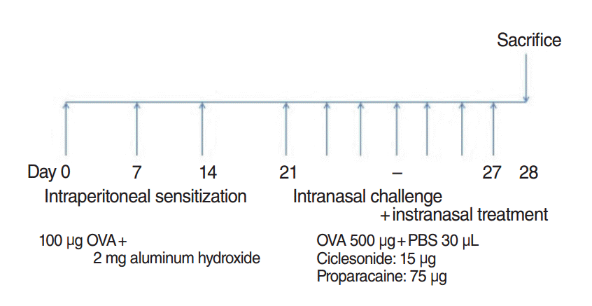
Schematic representation of the experimental allergic rhinitis model and treatment protocol. Briefly, on days 0, 7, and 14, mice were systemically sensitized by intraperitoneal administration of 100 μg of ovalbumin (OVA) mixed with 2 mg of aluminum hydroxide (Sigma-Aldrich) in 300 μL of phosphate-buffered saline (PBS) or PBS only (control group). During challenge, mice in the proparacaine (PPC) and ciclesonide (CIC) groups were treated intranasally with CIC at 15 μg/30 μL and PPC at 75 μg/30 μL, respectively, for 7 consecutive days.
Evaluation of nasal symptoms after final allergen challenge
Observers who were blinded to the mice groups counted how many times the mice sneezed or rubbed its nose during 10 minutes after the final allergen challenge.
Nasal mucosal tissue evaluation
Twenty-four hours after the final allergen challenge, mice were sacrificed. The mice were decapitated then the heads were fixed with 4% paraformaldehyde for 72 hours at 4°C. At room temperature, the heads were washed in running water then placed in Calci-Clear Rapid (National Diagnostics, Atlanta, GA, USA) for 72 hours for decalcification. The heads were then dehydrated by passing through a series of alcohol grades and then were placed in paraffin blocks. The paraffin blocks were then divided into 4-μm thick sections, and stained with hematoxylin and eosin stain, toluidine blue stain, and periodic acid Schiff’s stain (PAS).
Staining
Hematoxylin and eosin stain was used to quantify the number of eosinophils and aid the observer in describing the character of eosinophils in the lamina propria. Eosinophils were defined morphologically by the presence of 2-lobed nucleus and eosinophilic granules in the cytoplasm. Eosinophils were counted under a microscope at ×400 magnification [13]. Individuals who were blinded to the animals’ group assignments counted the number of eosinophils under a light microscope.
Toluidine blue staining was used to identify the mast cells. Sections were deparaffinized, stained with 0.5% toluidine blue solution, washed with water, differentiated with 0.5% glacial acetic acid solution until showing clear nuclei and cytoplasm granules. The mast cells were counted under light microscope. The number of mast cells was quantified at the mucosa of the nasal septum at ×400 magnification.
To observe goblet cell hyperplasia, PAS technique was used. First, the sections were deparaffinized and dehydrated. They were then stained with periodic acid and Schiff’s stain. Goblet cells stained with PAS stain were identified along the nasal septum mucosa with a light microscope at ×400 magnification.
Serum levels of total and OVA-specific IgE, IgG1, and IgG2a
Serum samples were obtained by 3,000 rpm centrifugation for 10 minutes. OVA-specific immunoglobulin (Ig) E, and IgG1 levels were measured by mouse anti-OVA IgE and anti-OVA IgG1 enzyme-linked immunosorbent assays (ELISA; Cayman Chemical, Ann Arbor, MI, USA). Anti-OVA IgG2a level was measured by Mouse Anti-OVA IgG2a antibody assay kit (Chondrex, Inc., Redmond, WA, USA).
Serum levels of interleukin-4 and interleukin-5
Serum levels of interleukin (IL)-4 and IL-5 were measured by ELISA kit (Quantikine ELISA kit, R&D systems, Minneapolis, MN, USA) according to instruction of manufacturer.
Statistical analysis
Statistical program IBM SPSS ver. 20.0 (IBM Co., Armonk, NY, USA) was used for statistical analysis. Measured parameters were expressed as means±standard deviation. Differences of nonparametric parameters between different groups were analyzed with Kruskal-Wallis test. If there were statistical significances, Bonferroni’s multiple comparison post-test was used to examine the differences between different group. The P-value less than 0.05 was considered to be statistically significant at a two-tailed test.
RESULTS
Nasal symptom score
The numbers of nasal rubbing were 5.11±3.62 in the control group, 25.78±15.66 in the AR group, 8.78±3.80 in the CIC group, and 9.89±5.04 in the PPC group. The numbers of sneezing were 1.44±1.51 in the control group, 21.33±7.48 in the AR group, 5.89±2.57 in the CIC group, and 8.56±5.41 in the PPC group. The number of nasal rubbing and sneezing in the AR group were significantly elevated compared to those in the control group. Nasal rubbing and sneezing were significantly reduced in the CIC and PPC groups compared to the AR group (both P<0.05) (Fig. 2).
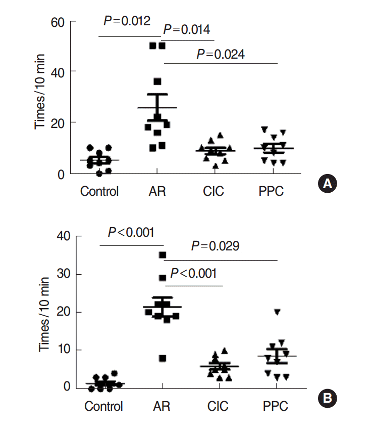
Nasal symptom score. Rubbing (A) and sneezing (B). AR, allergic rhinitis; CIC, ciclesonide; PPC, proparacaine.
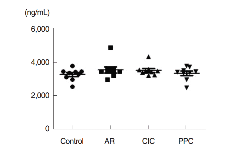
Serum OVA-specific IgE
Serum OVA-specific IgE level was considerably higher in the AR group, CIC group, and PPC group (7,965.19±5,285.72 ng/mL, 5,721.61±4,014.33 ng/mL, 6,995.88±3,385.95 ng/mL; P<0.001) than in the control group (77.57±69.50 ng/mL). No significant difference was observed between AR group, CIC group, and PPC group. Serum OVA-specific IgG1 level was also considerably higher in the AR group, CIC group, and PPC group (6,266,711.11±3,289,120.81 ng/mL, 4,828,688.89±1,642,127.15 ng/mL, 5,704,955.56±1,722,310.45 ng/mL; P=0.001) than in the control group (391,022.22±850,377.19 ng/mL). No significant difference was observed between AR group, CIC group and PPC group. Serum OVA-specific IgG2a level was also considerably higher in the AR group, CIC group, and PPC group (926.84±522.74 ng/mL, 924.14±424.89 ng/mL, 645.67±387.27 ng/mL; P<0.001) than in the control group (4.17±0.99 ng/mL). No significant difference was observed between AR group, CIC group, and PPC group (Fig. 3).
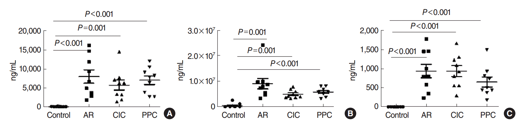
Levels of ovalbumin (OVA) specific immunoglobulin (Ig) E (A), OVA-specific IgG1 (B), and OVA-specific IgG2a (C) in the serum. OVA sensitization significantly increased OVA-specific IgE, OVA-specific IgG1, and OVA-specific IgG2, while intranasal treatment with CIC and PPC had no significant effect. AR, allergic rhinitis; CIC, ciclesonide; PPC, proparacaine.
Eosinophils counts in the lamina propria
Eosinophil counts were 2.78±1.64 in the control group, 47.33±13.58 in the AR group, 5.22±2.73 in the CIC group, and 5.89±3.33 in the PPC group. Eosinophil infiltration was observed at a significantly higher degree in the AR group in comparison with the control (P<0.001), CIC (P<0.001), and PPC (P<0.001) groups (Fig. 4).

Infiltration of eosinophils (arrows) in the nasal mucosa of BALB/c mice: (A) control group, (B) allergic rhinitis (AR) group, (C) ciclesonide (CIC) group, and (D) proparacaine (PPC) group (H&E, ×400). (E) Eosinophil counts in the nasal mucosa of each study group.
Goblet cell counts in the lamina propria
Goblet cell counts were 24.44±12.71 in the control group, 92.78±33.43 in the AR group, 28.11±10.12 in the CIC group, and 28.78±9.01 in the PPC group. Goblet cell infiltration was observed a significantly higher degree in the AR group in comparison with the control (P<0.001), CIC (P<0.001), and PPC (P<0.001) groups (Fig. 5).
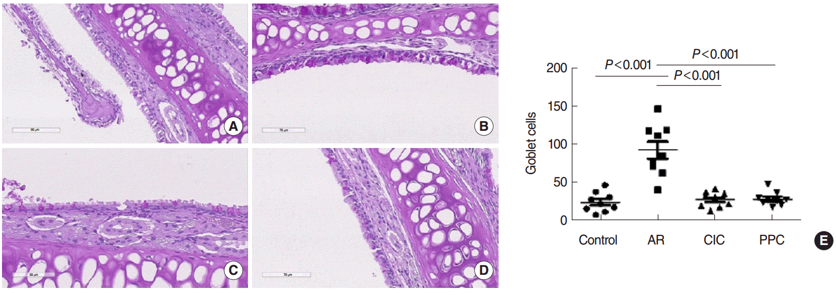
Infiltration of goblet cells in the nasal mucosa of BALB/c mice: (A) control group, (B) allergic rhinitis (AR) group, (C) ciclesonide (CIC) group, and (D) proparacaine (PPC) group (periodic acid Schiff staining, ×400). (E) Goblet cell counts in the nasal mucosa of each study group.
Mast cell counts in the lamina propria
Mast cell counts were 4.11±3.18 in the control group, 25.33±9.35 in the AR group, 6.44±3.09 in the CIC group, and 4.89±3.06 in the PPC group. Mast cells infiltration was observed at a significantly higher degree in the AR group than in the control (P<0.001), CIC (P<0.001), and PPC (P<0.001) groups (Fig. 6).
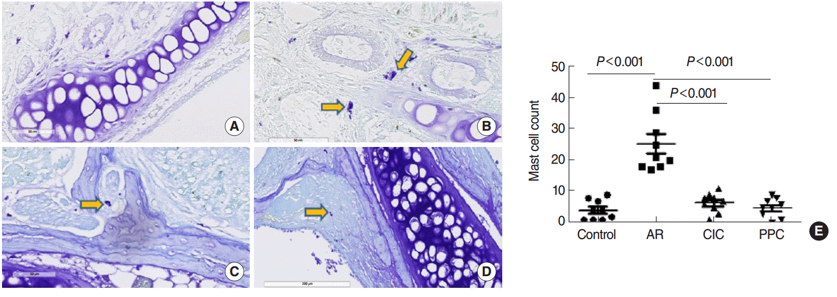
Infiltration of mast cells (arrows) in the nasal mucosa of BALB/c mice: (A) control group, (B) allergic rhinitis (AR) group, (C) ciclesonide (CIC) group, and (D) proparacaine (PPC) group (toluidine blue staining, ×400). (E) Mast cell counts in the nasal mucosa of each study group.
Serum levels of IL-4 and IL-5
Serum level of IL-4 was significantly lower in the CIC group and PPC group in comparison with AR group (both P<0.05). Serum IL-5 showed no significant difference between all groups (Fig. 7).
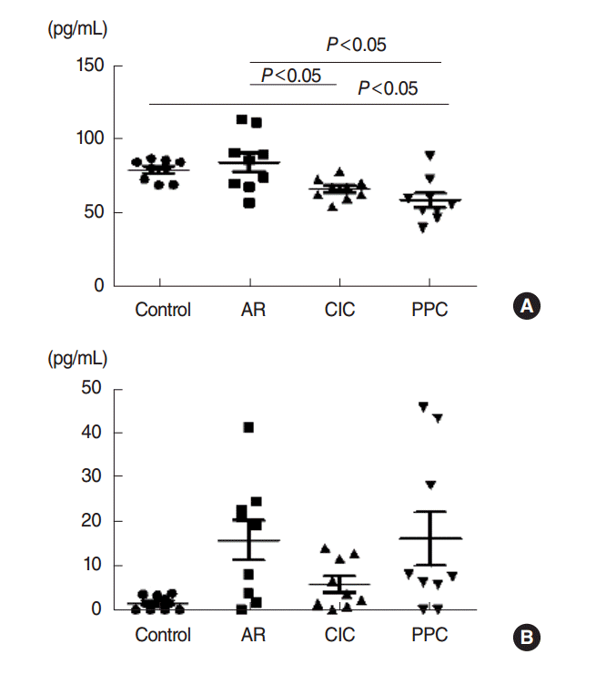
Effect of PPC on serum levels of Th2 cytokines, interleukin-4 (A), interleukin-5 (B). AR, allergic rhinitis; CIC, ciclesonide; PPC, proparacaine.
DISCUSSION
Lidocaine has emerged as a candidate drug for the treatment of refractory asthma [12,14-16]. A study found that airway inflammation can be reduced by nociceptor sensory neuron ablation, similar to the action of lidocaine [17]. The present study investigated the possibility that PPC, a derivative of lidocaine, exhibit anti-inflammatory effect in a mouse model of allergic rhinitis. We found that intranasal administration of PPC substantially reduced the allergic symptoms with inhibition of eosinophils, goblet cells, and mast cells infiltration in a similar degree with CIC.
We used PPC instead of lidocaine because PPC was readily available as a topical ophthalmic agent. We hypothesized that ophthalmic agent would be better absorbed by the nasal mucosa resulting in improved antiallergic effect.
Our study found that intranasal treatment of PPC inhibited eosinophil infiltration in the nasal mucosa of mouse of allergic rhinitis. This finding correlated with a previous study which used inhaled lidocaine in mouse model of asthma: lidocaine administration reduced subepithelial fibrosis in addition to peribronchial infiltration of eosinophil and neutrophil caused by OVA sensitization in lung tissue [6]. The exact mechanism has not been clearly identified but lidocaine is known to suppress the action of IL-3 and granulocyte-macrophage colony-stimulating factor resulting in reduced eosinophil survival and eosinophil superoxide production [5].
We found that intranasal administration of PPC inhibited goblet cell infiltration in the nasal mucosa of mouse model of allergic rhinitis. This finding was in concordance with a recent study which found that a lidocaine analog significantly inhibited OVA-induced mucus secretion in mice [18]. A different study found that topical application of lidocaine inhibited mucous secretion [19]. Our finding further supports previous findings in which lidocaine suppressed goblet cells.
Lastly, mast cell infiltration was reduced by treatment with PPC instillation in the nose of mouse model of allergic rhinitis. Such finding was also observed in a previous study which found lidocaine temporarily interfered with mast cell arrival [20]. A previous study discovered that lidocaine inhibited the allergen-evoked histamine release, in which mast cells play a crucial role [21,22].
With inhibition of eosinophils, goblet cells, and mast cells infiltration, the mouse treated with PPC showed decreased allergic nasal symptoms such as sneezing and nose rubbing.
Serum levels of IL-4 and IL-5 were examined to determine if serum cytokines reflected the decreased number of eosinophils in the nasal mucosa. As expected, serum level of IL-4 in PPC and CIC groups was significantly lower than AR group, but IL-5 did not show any significant difference. IL-4 and IL-5 are known to be representative cytokines of Th2 type immune response but we were not sure of the reason for such discrepancy between IL-4 and IL-5.
In our study, OVA specific IgE, OVA specific IgG1, and OVA specific IgG2a were significantly increased with intraperitoneal OVA sensitization which did not decrease after intranasal treatment with steroid or PPC. This might be different from previous studies which reported intranasal treatment with steroid or other therapeutic materials lowered blood levels of OVA specific IgE [13,23]. Interestingly, as mentioned above, serum levels of IL-4 were lower in the PPC and CIC groups in comparison with AR group. A recent study found that serum specific IgE may have a role in the generation of allergic inflammation but its role may not be crucial in a mouse model of AR [24]. Therefore, local specific IgE may be superior to serum specific IgE in representing the allergic inflammation at local nasal mucosa [24]. Further study will be needed to explain the discrepancy in the role between local and serum levels of specific IgE and Th2 cytokines.
Lastly, serum cortisol levels showed no significant difference between the groups. Serum cortisol levels reflect activity of the hypothalamus-pituitary-adrenal (HPA) axis which can be suppressed by exogenous steroid [25]. This finding might be explained by intranasal treatment with steroids had no systemic effect such as HPA axis suppression which might have been different from local nasal mucosa.
As a limitation of our study, we failed to measure levels of OVA specific IgE of nasal mucosa or nasal mucosal specific response including Th1, Th2, Th17, or innate cytokines which could have better reflected the therapeutic effect of PPC. However, we found increased levels of serum OVA specific IgE, OVA specific IgG1, and OVA specific IgG2a which showed that mouse model of allergic rhinitis had been properly established.
In conclusion, PPC, which is a derivative of lidocaine appears to be effective in treatment of mouse model of allergic rhinitis. This is the first study to demonstrate that a local anesthetic might have a therapeutic potential in treatment with allergic rhinitis. Further investigation of the anti-allergic mechanism of PPC seems to be worthy.
HIGHLIGHTS
▪ Antiallergic effect of proparacaine, a local anesthetic, was examined in a mouse model of allergic rhinitis.
▪ Proparacaine reduced nasal symptoms, sneezing, and nose rubbing.
▪ Proparacaine reduced eosinophil, mast cell, and goblet cell infiltration in the nasal mucosa.
Notes
No potential conflict of interest relevant to this article was reported.
Acknowledgements
The authors express our gratitude for the financial support of the Catholic Medical Center Research Foundation in the program, 2014. This study was funded by Outstanding Fellow Research Grant Award from the Korean Academy of Pediatric Allergy and Respiratory Disease in 2014.